 | Info
Sheets |
| | | | | | | | | | | | | | | | | | | | | | | | |
 | Out-
side |
| | | | |
|
| | | | |
Result : Searchterm 'Inversion Recovery' found in 13 terms [ ] and 30 definitions [ ] and 30 definitions [ ] ]
| | previous 16 - 20 (of 43) nextResult Pages : [1 2 3] [4 5 6 7 8 9] |  | |  | Searchterm 'Inversion Recovery' was also found in the following services: | | | | |
| |  |
| |
|
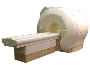
'Next generation MRI system 1.5T CHORUS developed by ISOL Technology is optimized for both clinical diagnostic imaging and for research development.
CHORUS offers the complete range of feature oriented advanced imaging techniques- for both clinical routine and research. The compact short bore magnet, the patient friendly design and the gradient technology make the innovation to new degree of perfection in magnetic resonance.'
Device Information and Specification
CLINICAL APPLICATION
Whole body
Spin Echo, Gradient Echo, Fast Spin Echo,
Inversion Recovery ( STIR, Fluid Attenuated Inversion Recovery), FLASH, FISP, PSIF, Turbo Flash ( MPRAGE ),TOF MR Angiography, Standard echo planar imaging package (SE-EPI, GE-EPI), Optional:
Advanced P.A. Imaging Package (up to 4 ch.), Advanced echo planar imaging package,
Single Shot and Diffusion Weighted EPI, IR/FLAIR EPI
STRENGTH
20 mT/m (Upto 27 mT/m)
| | | | | |
| | | | | |
| |
|
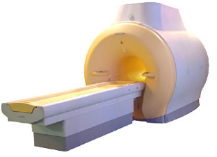
'MRI system is not an expensive equipment anymore.
ENCORE developed by ISOL Technology is a low cost MRI system with the advantages like of the 1.0T MRI scanner. Developed specially for the overseas market, the ENCORE is gaining popularity in the domestic market by medium sized hospitals.
Due to the optimum RF and Gradient application technology. ENCORE enables to obtain high resolution imaging and 2D/3D Angio images which was only possible in high field MR systems.'
- Less consumption of the helium gas due to the ultra-lightweight magnet specially designed and manufactured for ISOL.
- Cost efficiency MR system due to air cooling type (equivalent to permanent magnetic).
- Patient processing speed of less than 20 minutes.'
Device Information and Specification
CLINICAL APPLICATION
Whole body
CONFIGURATION
Short bore compact
| | | |
• View the DATABASE results for 'ENCORE 0.5T™' (2).
| | | | |
| | | | | |
| |
|
| | | |
• View the DATABASE results for 'Echo Time' (36).
| | | | |  Further Reading: Further Reading: | | Basics:
|
|
News & More:
| |
| |
| | | Searchterm 'Inversion Recovery' was also found in the following services: | | | | |
| | |
| |
|
Fat suppression is the process of utilizing specific MRI parameters to remove the deleterious effects of fat from the resulting images , e.g. with STIR, FAT SAT sequences, water selective (PROSET WATS - water only selection, also FATS - fat only selection possible) excitation techniques, or pulse sequences based on the Dixon method.
Spin magnetization can be modulated by using special RF pulses. CHESS or its variations like SPIR, SPAIR ( Spectral Selection Attenuated Inversion Recovery) and FAT SAT use frequency selective excitation pulses, which produce fat saturation.
Fat suppression techniques are nearly used in all body parts and belong to every standard MRI protocol of joints like knee, shoulder, hips, etc.

Image Guidance
Imaging of, e.g. the foot can induce bad fat suppression with SPIR/FAT SAT due to the asymmetric volume of this body part. The volume of the foot alters the magnetic field to a different degree than the smaller volume of the lower leg affecting the protons there. There is only a small band of tissue where the fat protons are precessing at the frequency expected, resulting in frequency selective fat saturation working only in that area. This can be corrected by volume shimming or creating a more symmetrical volume being imaged with water bags.
Even with their longer scan time and motion sensitivity, STIR (short T1/tau inversion recovery) sequences are often the better choice to suppress fat. STIR images are also preferred because of the decreased sensitivity to field inhomogeneities, permitting larger fields of views when compared to fat suppressed images and the ability to image away from the isocenter. See also Knee MRI.
Sequences based on Dixon turbo spin echo ( fast spin echo) can deliver a significant better fat suppression than conventional TSE/FSE imaging.
| | | | | |
• View the DATABASE results for 'Fat Suppression' (28).
| | | | | | Further Reading: | | Basics:
|
|
News & More:
| |
| |
| | | | | |
| |
|
A pulse sequence is a preselected set of defined RF and gradient pulses, usually repeated many times during a scan, wherein the time interval between pulses and the amplitude and shape of the gradient waveforms will control NMR signal reception and affect the characteristics of the MR images. Pulse sequences are computer programs that control all hardware aspects of the MRI measurement process.
Usual to describe pulse sequences, is to list the repetition time (TR), the echo time (TE), if using inversion recovery, the inversion time (TI) with all times given in milliseconds, and in case of a gradient echo sequence, the flip angle. For example, 3000/30/1000 would indicate an inversion recovery pulse sequence with TR of 3000 msec., TE of 30 msec., and TI of 1000 msec.
Specific pulse sequence weightings are dependent on the field strength, the manufacturer and the pathology.
See also Interpulse Times. | | | |
• View the DATABASE results for 'Pulse Sequence' (96).
| | |
• View the NEWS results for 'Pulse Sequence' (1).
| | | | | | Further Reading: | Basics:
|
|
News & More:
| |
| |
| | | | |
| | | |
|
| |
 | Look
Ups |
| |


