| Info
Sheets |
| | | | | | | | | | | | | | | | | | | | | | | | |
 | Out-
side |
| | | | |
|
| | | | |
Result : Searchterm 'brain imaging' found in 0 term [ ] and 2 definitions [ ] and 2 definitions [ ], (+ 19 Boolean[ ], (+ 19 Boolean[ ] results ] results
| | 1 - 5 (of 21) nextResult Pages : [1] [2 3 4 5] |  | |  | Searchterm 'brain imaging' was also found in the following services: | | | | |
| |  |
| |
|
Brain imaging, magnetic resonance imaging of the head or skull, cranial magnetic resonance tomography (MRT), neurological MRI - they describe all the same radiological imaging technique for medical diagnostic.
Magnetic resonance imaging of the human brain includes the anatomic description and the detection of lesions. Special techniques like diffusion weighted imaging, functional magnetic resonance imaging ( fMRI) and spectroscopy provide also information about the function and chemical metabolites of the brain.
MRI provides detailed pictures of brain and nerve tissues in multiple planes without obstruction by overlying bones. Brain MRI is the procedure of choice for most brain disorders. It provides clear images of the brainstem and posterior brain, which are difficult to view on a CT scan. It is also useful for the diagnosis of demyelinating disorders (disorders such as multiple sclerosis (MS) that cause destruction of the myelin sheath of the nerve).
With this noninvasive procedure also the evaluation of blood flow and the flow of cerebrospinal fluid (CSF) is possible. Different MRA methods, also without contrast agents can show a venous or arterial angiogram. MRI can distinguish tumors, inflammatory lesions, and other pathologies from the normal brain anatomy. However, MRI scans are also used instead other methods to avoid the dangers of interventional procedures like angiography (DSA - digital subtraction angiography) as well as of repeated exposure to radiation as required for computed tomography (CT) and other X-ray examinations.
A ( birdcage) bird cage coil achieves uniform excitation and reception and is commonly used to study the brain. Usually a brain MRI procedure includes FLAIR, T2 weighted and T1 weighted sequences in two or three planes. See also Fetal MRI, Fluid Attenuation Inversion Recovery ( FLAIR), Perfusion Imaging and High Field MRI. See also Arterial Spin Labeling. | | | | | | | | | | • Share the entry 'Brain MRI':    | | | | | | | | | |  Further Reading: Further Reading: | | Basics:
|
|
News & More:
|  |
MRI Reveals Significant Brain Abnormalities Post-COVID
Monday, 21 November 2022 by neurosciencenews.com | | |
Combining genetics and brain MRI can aid in predicting chances of Alzheimer's disease
Wednesday, 29 June 2022 by www.sciencedaily.com | | |
Roundup: How Even Mild COVID Can Affect the Brain; This Many Daily Steps Improves Longevity; and More
Friday, 11 March 2022 by baptisthealth.net | | |
A low-cost and shielding-free ultra-low-field brain MRI scanner
Tuesday, 14 December 2021 by www.nature.com | | |
Large International Study Reveals Spectrum of COVID-19 Brain Complications
Tuesday, 9 November 2021 by www.itnonline.com | | |
Brain MRI-Based Subtypes of MS Predict Disability Progression, Treatment Response
Thursday, 13 May 2021 by www.neurologyadvisor.com | | |
New MRI method improves detection of disease changes in the brain's network
Thursday, 11 June 2020 by www.compute.dtu.dk | | |
New NeuroCOVID Classification System Uses MRI to Categorize Patients
Friday, 12 June 2020 by www.diagnosticimaging.com | | |
New MRI technique can 'see' molecular changes in the brain
Thursday, 5 September 2019 by medicalxpress.com | | |
Talking therapy or medication for depression: Brain scan may help suggest better treatment
Monday, 27 March 2017 by www.newsnation.in | | |
MRI identifies brain abnormalities in chronic fatigue syndrome patients
Wednesday, 29 October 2014 by www.eurekalert.org | | |
MRIs Useful in Tracking Depression in MS Patients
Tuesday, 1 July 2014 by www.hcplive.com | | |
Contrast agent linked with brain abnormalities on MRI
Tuesday, 17 December 2013 by www.sciencecodex.com | | |
MRIs Reveal Signs of Brain Injuries Not Seen in CT Scans
Tuesday, 18 December 2012 by www.sciencedaily.com | | |
Iron Deposits in the Brain May Be Early Indicator of MS
Wednesday, 13 November 2013 by www.healthline.com | | |
Migraine Sufferers Have Thicker Brain Cortex
Tuesday, 20 November 2007 by www.medicalnewstoday.com |
|
| |
| | | | | |
| |
|
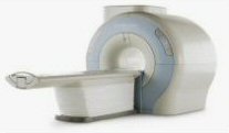
From GE Healthcare;
The Signa HDx MRI system is GE's leading edge whole body magnetic resonance scanner designed to support high resolution, high signal to noise ratio, and short scan times.
Signa HDx 3.0T offers new technologies like ultra-fast image reconstruction through the new XVRE recon engine, advancements in parallel imaging algorithms and the broadest range of premium applications. The HD applications, PROPELLER (high-quality brain imaging extremely resistant to motion artifacts), TRICKS (contrast-enhanced angiographic vascular lower leg imaging), VIBRANT (for breast MRI), LAVA (high resolution liver imaging with shorter breath holds and better organ coverage) and MR Echo (high-definition cardiac images in real time) offer unique capabilities.
Device Information and Specification CLINICAL APPLICATION Whole body
CONFIGURATION Compact short bore SE, IR, 2D/3D GRE, RF-spoiled GRE, 2DFGRE, 2DFSPGR, 3DFGRE, 3DFSPGR, 3DTOFGRE, 3DFSPGR, 2DFSE, 2DFSE-XL, 2DFSE-IR, T1-FLAIR, SSFSE, EPI, DW-EPI, BRAVO, Angiography: 2D/3D TOF, 2D/3D phase contrast vascular IMAGING MODES Single, multislice, volume study, fast scan, multi slab, cine, localizer H*W*D 240 x 2216,6 x 201,6 cm POWER REQUIREMENTS 480 or 380/415, 3 phase ||
COOLING SYSTEM TYPE Closed-loop water-cooled grad. | | | | | |
| | | | |  |
| |
|
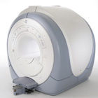
Device Information and Specification
CLINICAL APPLICATION
Whole body
SE, IR, FSE, FIR, GE, SG, BASG, PBSG, PCIR, DWI, Radial, Angiography: TOF, FLUTE (Fluoro-triggered bolus MRA), Time-resolved MRA
IMAGING MODES
Single, multislice, volume study
Level Range: -2,000 to +4,000
POWER REQUIREMENTS
208/220/240 V, single phase
| | | |
• View the DATABASE results for 'Echelon™ 1.5T' (2).
| | |
• View the NEWS results for 'Echelon™ 1.5T' (3).
| | | | | | Further Reading: | Basics:
|
|
| |
| | | Searchterm 'brain imaging' was also found in the following services: | | | | |
| | |
| |
|
Contact Information
MAIL
Bornhop Research Group
MS 1061 Dept of Chemistry and Biochemistry
Texas Tech University
Lubbock, TX 79409
USA
PHONE
Office Phone: +1-806-742-3142
Lab Phone: +1-806-742-3152
| | | | | |
| | | | | |
| |
|
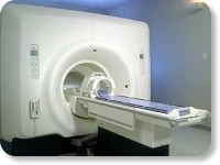
(Signa VH/i 3.0T)
With GE Healthcare
leading-edge technology in ultra-high-field imaging. The 3 T VH/i provides a platform for advanced applications in radiology, cardiology, psychology and psychiatry. Real-time image processing lets you acquire multislice whole brain images and map brain functions for research or surgical planning. And the 3 T Signa VH/i is flexible enough to provide clinicians with high performance they require. It can provide not only outstanding features in brain scanning and neuro-system research, but also a wide range of use in scanning breasts, extremities, the spine and the cardiovascular systems.
Device Information and Specification CLINICAL APPLICATION Whole body
T/R quadrature head, T/R quadrature body, T/R phased array extremity (opt) SE, IR, 2D/3D GRE, FGRE, RF-spoiled GRE, FSE, Angiography: 2D/3D TOF, 2D/3D phase contrast vascular IMAGING MODES Single, multislice, volume study, fast scan, multi slab, cine, localizer 100 Images/sec with Reflex100 MULTISLICE 100 Images/sec with Reflex100 2D 0.5-100mm in 0.1mm incremental 128x512 steps 32 phase encode H*W*D 260cm x 238cm x 265cm POWER REQUIREMENTS 480 or 380/415, 3 phase ||
COOLING SYSTEM TYPE Closed-loop water-cooled grad. Less than 0.14 L/hr liquid He | | | |
• View the DATABASE results for 'Signa 3.0T™' (2).
| | | | |
| | | | |
| | | 1 - 5 (of 21) nextResult Pages : [1] [2 3 4 5] |
| |
|
| |
 | Look
Ups |
| |