 | Info
Sheets |
| | | | | | | | | | | | | | | | | | | | | | | | |
 | Out-
side |
| | | | |
|
| | | | | |  |
Result : Searchterm 'mrcp' found in 0 term [ ] and 13 definitions [ ] and 13 definitions [ ] ]
| | 1 - 5 (of 13) nextResult Pages : [1 2 3] | | |  | Searchterm 'mrcp' was also found in the following services: | | | | |
| |  |
| |
|
(SENSE) A MRI technique for relevant scan time reduction. The spatial information related to the coils of a receiver array are utilized for reducing conventional Fourier encoding. In principle, SENSE can be applied to any imaging sequence and k-space trajectories. However, it is particularly feasible for Cartesian sampling schemes. In 2D Fourier imaging with common Cartesian sampling of k-space sensitivity encoding by means of a receiver array enables to reduce the number of Fourier encoding steps.
SENSE reconstruction without artifacts relies on accurate knowledge of the individual coil sensitivities. For sensitivity assessment, low-resolution, fully Fourier-encoded reference images are required, obtained with each array element and with a body coil.
The major negative point of parallel imaging techniques is that they diminish SNR in proportion to the numbers of reduction factors.
R is the factor by which the number of k-space samples is reduced. In standard Fourier imaging reducing the sampling density results in the reduction of the FOV, causing aliasing. In fact, SENSE reconstruction in the Cartesian case is efficiently performed by first creating one such aliased image for each array element using discrete Fourier transformation (DFT).
The next step then is to create a full-FOV image from the set of intermediate images. To achieve this one must undo the signal superposition underlying the fold-over effect. That is, for each pixel in the reduced FOV the signal contributions from a number of positions in the full FOV need to be separated. These positions form a Cartesian grid corresponding to the size of the reduced FOV.
The advantages are especially true for contrast-enhanced MR imaging such as
dynamic liver MRI (liver imaging) ,
3 dimensional magnetic resonance angiography (3D MRA), and magnetic resonance cholangiopancreaticography ( MRCP).
The excellent scan speed of SENSE allows for acquisition of two separate sets of hepatic MR images within the time regarded as the hepatic arterial-phase (double arterial-phase technique) as well as that of multidetector CT.
SENSE can also increase the time efficiency of spatial signal encoding in 3D MRA. With SENSE, even ultrafast (sub second) 4D MRA can be realized.
For MRCP acquisition, high-resolution 3D MRCP images can be constantly provided by SENSE. This is because SENSE resolves the presence of the severe motion artifacts due to longer acquisition time. Longer acquisition time, which results in diminishing image quality, is the greatest problem for 3D MRCP imaging.
In addition, SENSE reduces the train of gradient echoes in combination with a faster k-space traversal per unit time, thereby dramatically improving the image quality of single shot echo planar imaging (i.e. T2 weighted, diffusion weighted imaging). | | | | | | • Share the entry 'Sensitivity Encoding':    | | | | | | | | | |  Further Reading: Further Reading: | News & More:
|
|
| |
| | | | | |
| |
|
Categories of negative oral contrast agents:
Negative oral contrast media are usually based on superparamagnetic particles and act by inducing local field inhomogeneities, which results in shortening of both T1 and T2 relaxation times. Superparamagnetic contrast agents have predominant T2 weighted effects.
Biphasic contrast media are agents that have different signal intensities on different sequences, depending on the concentration at which they are used.
Suitable materials for oral contrast agents should have little or no absorption by the stomach or intestines, complete excretion, no motion or susceptibility artifacts, affordability, and uniform marking of the gastrointestinal tract.
Benefits of negative oral contrast agents are the reduction of ghosting artifacts caused by the lack of signal. Superparamagnetic iron oxides produce also in low concentrations a noticeable signal loss; but can generate susceptibility artifacts especially in gradient echo sequences. Perfluorochemicals do not dilute in the bowel because they are not miscible with water.
High cost, poor availability, and limited evaluations of side effects are possible disadvantages.
Negative oral contrast agents are used e.g., in MRCP, where the ingestion of 600-900 ml of SPIO cancels out the signal intensity of the lumen (in addition after the injection of a gadolinium-based contrast medium, the enhancement of the inflammatory tissues is clearer seen), and in MR abdominal imaging of Crohn's disease in combination with mannitol.

| | | |
• View the DATABASE results for 'Negative Oral Contrast Agents' (7).
| | | | | | Further Reading: | Basics:
|
|
| |
| | | | | |
| |
|
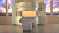
From Hitachi Medical Systems America Inc.;
the AIRIS II, an entry in the diagnostic category of open MR systems, was designed by Hitachi
Medical Systems America Inc. (Twinsburg, OH, USA) and Hitachi Medical Corp. (Tokyo) and is manufactured by the Tokyo branch. A 0.3 T field-strength magnet and phased array coils deliver high image quality without the need for a tunnel-type high-field system, thereby significantly improving patient comfort not only for claustrophobic patients.
Device Information and Specification
CLINICAL APPLICATION
Whole body
QD Head, MA Head and Neck, QD C-Spine, MA or QD Shoulder, MA CTL Spine, QD Knee, Neck, QD TMJ, QD Breast, QD Flex Body (4 sizes), Small and Large Extrem., QD Wrist, MA Foot and Ankle (WIP), PVA (WIP)
SE, GE, GR, IR, FIR, STIR, FSE, ss-FSE, FLAIR, EPI -DWI, SE-EPI, ms - EPI, SSP, MTC, SARGE, RSSG, TRSG, MRCP, Angiography: CE, 2D/3D TOF
IMAGING MODES
Single, multislice, volume study
TR
SE: 30 - 10,000msec GE: 20 - 10,000msec IR: 50 - 16,700msec FSE: 200 - 16,7000msec
TE
SE : 10 - 250msec IR: 10 -250msec GE: 5 - 50 msec FSE: 15 - 2,000
0.05 sec/image (256 x 256)
2D: 2 - 100 mm; 3D: 0.5 - 5 mm
Level Range: -2,000 to +4,000
POWER REQUIREMENTS
208/220/240 V, single phase
COOLING SYSTEM TYPE
Air-cooled
2.0 m lateral, 2.5 m vert./long
| | | |
• View the DATABASE results for 'AIRIS II™' (2).
| | | | |
| | | Searchterm 'mrcp' was also found in the following services: | | | | |
| | |
| |
|
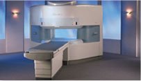
From Hitachi Medical Systems America, Inc.;
the AIRIS made its debut in 1995. Hitachi followed up with the AIRIS II system, which has proven equally successfully. 'All told, Hitachi has installed more than 1,000 MRI systems in the U.S., holding more than 17 percent of the total U.S. MRI installed base, and more than half of the installed base of open MR systems,' says Antonio Garcia, Frost and Sullivan industry research analyst.
Now Altaire employs a blend of innovative Hitachi features called VOSI™ technology, optimizing each sub-system's performance in concert with the
other sub-systems, to give the seamless mix of high-field performance
and the patient comfort, especially for claustrophobic patients, of open MR systems.
Device Information and Specification
CLINICAL APPLICATION
Whole body
DualQuad T/R Body Coil, MA Head, MA C-Spine, MA Shoulder, MA Wrist, MA CTL Spine, MA Knee, MA TMJ, MA Flex Body (3 sizes), Neck, small and large Extremity, PVA (WIP), Breast (WIP), Neurovascular (WIP), Cardiac (WIP) and MA Foot//Ankle (WIP)
SE, GE, GR, IR, FIR, STIR, ss-FSE, FSE, DE-FSE/FIR, FLAIR, ss/ms-EPI, ss/ms EPI- DWI, SSP, MTC, SE/GE-EPI, MRCP, SARGE, RSSG, TRSG, BASG, Angiography: CE, PC, 2D/3D TOF
IMAGING MODES
Single, multislice, volume study
TR
SE: 30 - 10,000msec GE: 3.6 - 10,000msec IR: 50 - 16,700msec FSE: 200 - 16,7000msec
TE
SE : 8 - 250msec IR: 5.2 -7,680msec GE: 1.8 - 2,000 msec FSE: 5.2 - 7,680
0.05 sec/image (256 x 256)
2D: 2 - 100 mm; 3D: 0.5 - 5 mm
Level Range: -2,000 to +4,000
COOLING SYSTEM TYPE
Water-cooled
3.1 m lateral, 3.6 m vertical
| | | |
• View the DATABASE results for 'Altaire™' (2).
| | | | | | Further Reading: | News & More:
|
|
| |
| | | | | |
| |
|
Diamagnetism occurs only by a substance in the presence of an externally applied magnetic field. Diamagnetic contrast agents are complexes in which the metal ion (e.g., Zn, Bi and Ca) is diamagnetic.
Potential diamagnetic materials in gastrointestinal MRI:
A suspension of clay minerals (Kaopectate with kaolin, a common over the counter drug) can be used as a negative oral contrast agent caused by the diamagnetic properties.
By using this preparation as a gastrointestinal contrast agent e.g., in pancreas MRI or MRCP, the absence of signal is clearly visible in the stomach and duodenum.
Barium sulfate commonly used as an X-ray contrast agent has also been tested for use in abdominal imaging. The diamagnetic properties of the barium particles are caused by a susceptibility effect around them, the resulting signal loss is strengthening by a replacement of water protons with barium.
See also Diamagnetism. | | | |
• View the DATABASE results for 'Gastrointestinal Diamagnetic Contrast Agents' (7).
| | | | |
| | | | |
| | |
| | | 1 - 5 (of 13) nextResult Pages : [1 2 3] |
| |
|
| |
 | Look
Ups |
| |


