|
Magnetic
Resonance -
Technology
Information
Portal |
Welcome to MRI Technology • • |
|
|
 | Info
Sheets |
| | | | | | | | | | | | | | | | | | | | | | | | |
 | Out-
side |
| | | | |
|
| | | | | |
| Result: Searchterm 'Anterior'
found in 5 messages |
| Result Pages: [1] |
More Results:  Database (7) News Service (5) Database (7) News Service (5) |
|
|
jung kang
Fri. 29 Dec.23,
02:10
[Start of:
'Chronic insufficiency of the PCL is suspected.'
0 Reply]
 Category: Category:
Funktional MRI
|
| Chronic insufficiency of the PCL is suspected. |
Hello,
I'm experiencing a specific issue with my knee and would appreciate any insights. When I straighten my knee, I feel a friction sensation at the junction of the tibia behind the knee. This sensation of friction is very close to the area of pain, which seems to be deep and difficult to pinpoint accurately.
Here's a summary of my medical reports and symptoms:
First MRI Report Findings:
Chronic damage to the posterior cruciate ligament.
Patellofemoral wear and tear.
I suspect these findings are related to my symptoms.
Additional Information:
A CT scan showed swelling in the anterior cruciate ligament.
Pain Description:
The pain starts from the middle of the knee joint and gradually spreads to the right popliteal fossa (back of the knee).
I experience this pain continuously for 24 hours.
Other Symptoms:
Along with the onset of these symptoms, I've noticed an unusually large amount of hair loss, which I suspect might be due to ongoing inflammation.
I'm seeking advice or insights from anyone who might have had similar experiences or from medical professionals who can shed some light on these symptoms and the MRI findings.
Thank you in advance for your help!
2023
- https://www.dicomlibrary.com/meddream/?study=1.3.6.1.4.1.44316.6.102.1.202312232236683.4754602657439816271065
2021
- https://www.dicomlibrary.com/meddream/?study=1.3.6.1.4.1.44316.6.102.1.20231225201235919.34251024914768354353
|
| |  Reply to this thread Reply to this thread
(login or register first) | |
Arnold Somereville
Sun. 23 Apr.17,
18:06
[Start of:
'GE 8 channel Body Coil Artifact'
2 Replies]
Category:
Artifacts
|
| GE 8 channel Body Coil Artifact |
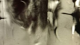
Have a GE 1.5T HDxt. Problem: experiencing symmetric artifacts in abdominal images. Artifact seen in most anterior images: Some are rectangular in appearance, another reminds me of a plastic knife. All patients are changed in gowns/pants prior to entry into MRI suite. The body coil in question has been changed 3 times but the artifact remains. I suspect this is a hardware reconstruction issue, however, the engineer thinks it is operator error. I have run this sequence with and without options. This is an SSFSE, breath hold sequence. Has anyone encountered this issue?
 MRI Abdomen 8 channel body coil MRI Abdomen 8 channel body coil
|
| | View the whole thread | Reply to this thread
(login or register first) | | |
|
Bob Smtih
Thu. 30 Oct.14,
18:41
[Start of:
'What is this white "cloud" on the right side of this MRI?'
0 Reply]
Category:
General
|
| What is this white "cloud" on the right side of this MRI? |
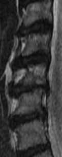
The following was noted:c2/c3 normal c3/c4 Very minimal anterior ostepphytic ridging is present. There is no spinal stenosis or neural foraminal narrowing. C4/c5 Very minimal anterior ostepphytic ridging is noted. There is no spinal stenosis or neural foraminal narrowing. c5/c6 There is a circumferential disc osteophyte complex present. There is mild central spinal stenosis. The AP diameter of the spinal canal is narrowed to 9mm. The dorsal and ventral CSF spaces remain patent. There is mild to moderate neural foraminal narrowing. c6/c7 a circumferential disc osteophyte complex present. There is mild central spinal stenosis. AP diameter of the spinal canal is narrowed to 9mm. The dorsal and ventral CSF spaces remain patent. There is moderate to severe neural foraminal narrowing. c7/t1 Normal. AP diameter of the spinal canal is 12 mm My question is C5 looks like it is chewed up; weird shading. Does it appear to be more damaged than the report states? Also there seems to be a white "cloud" that goes into the left side of multiple disks. What is the white "cloud"? c3 through c7 are in the image
MRI SAG T2
|
| | Reply to this thread
(login or register first) | |
Mike Parks
Fri. 6 Apr.07,
18:53
[Start of:
'Need Help'
0 Reply]
Category:
Applications and Examinations
|
| Need Help |
I had an MRI yesterday and I have alot of metal in my neck due to extensive surgery such as anterior fusion from c4 to c7. Yesterdays results indicate I have atrophy of the spinal cord from C4-5 to C7 with a thin cord and high signal compatible with myelomalacia. Herniation of C3-$ that compresses the spinal cord. Central protrusion of the C2-3 and C7-T1 discs. My question is. My neck burns as if on fire inside. Is this something that will go away? I have and do have severe pain 24/7 but I've never had this kind of pain. If possible please email me at oceansmove07@aol.com if you know what's happening. Thank you for any help you may have and I wish everyone a lighter today, a brighter tomorrow and a future filled with joy and happiness.
|
| | Reply to this thread
(login or register first) | |
GONZALO SOLIS
Tue. 22 Nov.05,
05:35
[Start of:
'usmarine2tim2'
0 Reply]
Category:
Basics and Physics
|
| usmarine2tim2 |
Hello to all my fellow MR Technologists and / or students:
I have a few questions that I would greatly appreciate if someone could help me answer. The dead line is THIS Friday 25th 1000AM morning so please reply STAT
Please e mail me at USMARINE2TIM2@YAHOO.COM
1. . Breath hold duration of less than _________are
recommended when dealing with MR Arenal artery
scan?
a. 30sec
b. 25sec
c. 18sec
d. 8
2. . In renal angiography, what anatomical
landmark does the 3d slab need to cover just
anterior to?
a. Adrenal glands
b. Hepatic vein
c. Portal vein
d. Abdominal aorta
3. .What makes MRI a better choice than DSA for evaluation of a bilateral renal artery stenoses at the orgins of the renal arteries?
a. It is a noisier examination
b. The contrast used is less nephrotoxic than DSA
contrast
c. MRI has higher special resolution than DSA
4. Signal loss will result when further maturation of the clot produces which one of the following?
a. methemoglobin
b. hemosiderin
c. phagocytosis.
d. hemoglobin
5. .For cine gradient echo imaging with true FISP, which of the following statements is TRUE?
a. .images have better image contrast and acquisitions are faster than conventional cine gradient echo imaging
b. cine true FISP images can be performed on any MRI unit
c. to make true FISD images, intravenous gadolinium contrast is needed
d. .true FISP images cannot be used for breath- hold acquisitions because the sequence is too slow
6. Which one of the following statements is NOT
true about phase contrast imaging?
a. .phase contrast imaging is useful for measuring blood flow
b. .phase contrast images are ECG gated
c. .phase contrast images can be performed on most
scanners with cardiac imaging capabilities
d. phase contrast images required
intravenous gadolinium contrast
Thank you so much
Gonzalo RT (R) --future-Ã MR
God Bless
|
| | Reply to this thread
(login or register first) | |
| |
| | Result Pages : [1] | |
|
| |
 | Look
Ups |
| |
|
MR-TIP.com uses cookies! By browsing MR-TIP.com, you agree to our use of cookies. | | | [last update: 2024-02-26 03:41:00] |
|
|