Magnetic Resonance - Technology Information Portal Welcome to MRI Technology
Info
  Sheets

Out-
      side
 

New acceleration techniques will :
reduce scan times 
cause artifacts 
increase expenses 
be useful if you have a lot of experience 
doesn't do much 
never heard of 


MRI Images
  Images Overview
 
 PCA-MRA 3D Brain Venography Colored MIP  
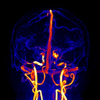
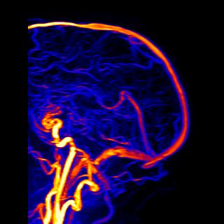
Phase contrast angiography (PCA) uses magnetic gradient characteristics and the developed phase shift of flowing blood in order to encode the blood flow velocity. Intracranial venous PCA-MRA (MR venography) provides the visualization of the dural venous sinuses (superior sagittal, transverse, straight and sigmoid sinuses), the larger deep cerebral (cavernous sinus) and cortical veins without the need for a contrast agent. The projection shows a transverse view of brain vessels.
 
with enhanced brightness and contrast
 
The magnetic resonance angiography (MRA) images are reconstructed in maximum intensity projection (MIP) and colored with the MRIcro postprocessing tool. Blue colors indicate small veins with slow flow. The venous sinuses are red colored; brighter colors indicate vessels with a faster blood flow velocity like the carotid arteries. The view shows a coronal processed MIP.
 
with enhanced brightness and contrast
 
The lateral view shows the sagittal brain sinus. 3D volume PCA provides a better image quality than 2D single-slice acquisition. PCA is more sensitive for slow flows (which are saturated in time of flight (TOF) MRA) and provides an excellent background suppression. Typical velocity encoding (VENC) parameters in brain MRI phase contrast sequences are 15-30 cm/sec for veins and 40-50 cm/sec for arteries.
 
with enhanced brightness and contrast
 
The PCA technique is based on a gradient echo sequence (for example: repetition time 10 ms, echo time TE 4.6 ms, flip angle 15, matrix 256, velocity encoding (VENC) 20 cm/sec, 1.5 Tesla). A parallel imaging technique is used to reduce acquisition time. Magnitude and velocity phase contrast MR images are acquired and processed by MIP. MR venography of the brain is used for the detection of venous thrombosis. PCA can also be helpful after application of a contrast medium in cases of wrong bolus timing or incorrect contrast enhanced magnetic resonance angiography acquisition.
 
with enhanced brightness and contrast
 
 
Related Images
 • Brain MRI Inversion Recovery
 • Brain MRI Coronal FLAIR 001
 • Brain MRI Sagittal T1 001
 • Brain MRI Transversal T1 001
 • Brain MRI Transversal T1 002
 • Brain MRI Transversal T2 001
 • Brain MRI Transversal T2 002
 • Brain MRI Transversal T2 003
 • Brain MRI Images Axial T2
 • Brain MRI Images T1
 • TOF-MRA Circle of Willis Inverted MIP
Related Sliders
 • MRI of the Skull Base
 • MRI of the Brain Stem with Temoral Bone and Auditory System
 • Circle of Willis, Time of Flight, MIP


Relevant Database Entries
 • 3 Dimensional Magnetic Resonance Angiography
 • Bipolar Gradient Pulse
 • Brain MRI
 • Contrast Enhanced MR Venography
 • Flow Effects
 • Magnetic Resonance Angiography MRA
 • Maximum Intensity Projection
 • Phase Contrast Angiography
 • Phase Contrast Sequence
 • Phase Shift
 • Postprocessing
 • Velocity Encoding
 
  Next Image - Anatomic Imaging of the Lungs top
In the beginning the Universe was created. This has made a lot of people very angry and been widely regarded as a bad move.
- Douglas Adams
 
Share This Page
FacebookTwitterLinkedIn

MR-TIP    
Community   
User
Pass
Forgot your UserID/Password ?    


Look
      Ups


MR-TIP.com uses cookies! By browsing MR-TIP.com, you agree to our use of cookies.

Magnetic Resonance - Technology Information Portal
Member of SoftWays' Medical Imaging Group - MR-TIP • Radiology-TIP • Medical-Ultrasound-Imaging • 
Copyright © 2003 - 2024 SoftWays. All rights reserved. [ 19 March 2024]
Terms of Use | Privacy Policy | Advertising
 [last update: 2024-02-26 03:41:00]