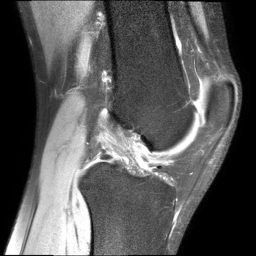
This interactive image series consists of thin slices of a proton density weighted sequence with fat suppression. The MR images show the anatomy of the knee in sagittal view from lateral (slice 1) to medial (slice 27). The used MRI scan shows high contrast between different structures of interest (e.g. bones, menisci, cruciate ligaments and cartilage).
The slices 14-16 show ghosting artifacts of the popliteal artery due to anterior/posterior phase encoding.
Remember not only to say the right thing in the right place, but far more
difficult still, to leave unsaid the wrong thing at the tempting moment. -
Benjamin Franklin