 | Info
Sheets |
| | | | | | | | | | | | | | | | | | | | | | | | |
 | Out-
side |
| | | | |
|
| | | | |
Result : Searchterm '3t' found in 1 term [ ] and 16 definitions [ ] and 16 definitions [ ] ]
| | previous 11 - 15 (of 17) nextResult Pages : [1] [2 3 4] |  | |  | Searchterm '3t' was also found in the following services: | | | | |
| |  |
| |
|
Knee MRI, with its high soft tissue contrast is one of the main imaging tools to depict knee joint pathology. MRI allows accurate imaging of intra-articular structures such as ligaments, cartilage, menisci, bone marrow, synovium, and adjacent soft tissue.
Knee exams require a dedicated extremity coil, providing a homogenous imaging volume and high SNR to ensure best signal coverage.
A complete knee MR examination includes for example sagittal and coronal T1 weighted, and proton density weighted pulse sequences +/- fat saturation, or STIR sequences. For high spatial resolution, maximal 4 mm thick slices with at least an in plane resolution of 0.75 mm and small gap are recommended. To depict the anterior cruciate ligament clearly, the sagittal plane has to be rotated 10 - 20° externally (parallel to the medial border of the femoral condyle). Retropatellar cartilage can bee seen for example in axial T2 weighted gradient echo sequences with Fatsat. However, the choice of the pulse sequences is depended of the diagnostic question, the used scanner, and preference of the operator.
Diagnostic quality in knee imaging is possible with field strengths ranging from 0.2 to 3T. With low field strengths more signal averages must be measured, resulting in increased scan times to provide equivalent quality as high field strengths.
More diagnostic information of meniscal tears and chondral defects can be obtained by direct magnetic resonance arthrography, which is done by introducing a dilute solution of gadolinium in saline (1:1000) into the joint capsule. The knee is then scanned in all three planes using T1W sequences with fat suppression. For indirect arthrography, the contrast is given i.v. and similar scans are started 20 min. after injection and exercise of the knee.
Frequent indications of MRI scans in musculoskeletal knee diseases are: e.g., meniscal degeneration and tears, ligament injuries, osteochondral fractures, osteochondritis dissecans, avascular bone necrosis and rheumatoid arthritis. See also Imaging of the Extremities and STIR. | | | | | | | | | | | | |  Further Reading: Further Reading: | | Basics:
|
|
News & More:
| |
| |
| | | | | |
| |
|
From Siemens Medical Systems;
Received FDA clearance in 2013.
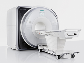
The MAGNETOM Prisma is the 3T PowerPack for exploration that offers most demanding clinical and research challenges of today and the future. The latest parallel transmit technology, TimTX TrueShape, enables zooming into specific body regions for enhanced image quality. Furthermore, the Tim 4G integrated coil technology offers remarkable imaging flexibility and supports complex
examinations across the whole body.
Onsite upgrades to the MAGNETOM Prisma for customers who have already installed the 3 Tesla MAGNETOM Trio are possible.
Device Information and Specification
CLINICAL APPLICATION
Whole Body
CONFIGURATION
Ultra-short bore
Head, spine, torso/ body coil, neurovascular, cardiac, neck, shoulder, knee, wrist, foot//ankle and multi-purpose flex coils. Peripheral vascular, breast, shoulder.
CHANNELS (min. / max. configuration)
64, 128
MAGNET WEIGHT (gantry included)
13000 kg
DIMENSION H*W*D (gantry included)
173 x 230 x 222 cm
Passive, active; first order,
second order
POWER REQUIREMENTS
380 / 400 / 420 / 440 / 460 / 480 V, 3-phase + ground;
| | | | | |
| | | | | |
| |
|

Odin Medical Technologies, Ltd. was founded in Israel, in 1996. Odin Medical Technologies, Inc. was established in early 2000 in the United States as a wholly owned subsidiary. Odin Medical Technologies develops and manufactures intraoperative magnetic resonance imaging ( MRI) image guidance systems designed to improve neurosurgical interventions with a special focus on minimally invasive procedures.
As of July 25, 2006, Odin Medical Technologies, Ltd. was acquired by Medtronic, Inc.
MRI Scanners:
| | | |
• View the DATABASE results for 'Odin Medical Technologies, Inc.' (2).
| | | | |
| | | Searchterm '3t' was also found in the following services: | | | | |
| | |
| |
|
The interactions between the most tested heart valve prostheses and the magnetic field of MRI devices are of no significance. However, there are many different types of heart valve prostheses and in the particular case their MRI safety should be checked.
Prosthetic heart valves, depending on the type and the material, are not necessarily considered to be dangerous in fields up to 3T. Patients should not undergo high field MRI examinations if valve dehiscence is clinically suspected. | | | |
• View the DATABASE results for 'Prosthetic Heart Valves' (2).
| | | | | | Further Reading: | Basics:
|
|
News & More:
| |
| |
| | | | | |
| |
|
From GE Healthcare;
The Signa HDx MRI system is GE's leading edge whole body magnetic resonance scanner designed to support high resolution, high signal to noise ratio, and short scan times.
Signa HDx 3.0T offers new technologies like ultra-fast image reconstruction through the new XVRE recon engine, advancements in parallel imaging algorithms and the broadest range of premium applications. The HD applications, PROPELLER (high-quality brain imaging extremely resistant to motion artifacts), TRICKS (contrast-enhanced angiographic vascular lower leg imaging), VIBRANT (for breast MRI), LAVA (high resolution liver imaging with shorter breath holds and better organ coverage) and MR Echo (high-definition cardiac images in real time) offer unique capabilities.
Device Information and Specification CLINICAL APPLICATION Whole body
CONFIGURATION Compact short bore SE, IR, 2D/3D GRE, RF-spoiled GRE, 2DFGRE, 2DFSPGR, 3DFGRE, 3DFSPGR, 3DTOFGRE, 3DFSPGR, 2DFSE, 2DFSE-XL, 2DFSE-IR, T1-FLAIR, SSFSE, EPI, DW-EPI, BRAVO, Angiography: 2D/3D TOF, 2D/3D phase contrast vascular IMAGING MODES Single, multislice, volume study, fast scan, multi slab, cine, localizer H*W*D 240 x 2216,6 x 201,6 cm POWER REQUIREMENTS 480 or 380/415, 3 phase ||
COOLING SYSTEM TYPE Closed-loop water-cooled grad. | | | | | |
| | | | |
| | | |
|
| |
 | Look
Ups |
| |


