 | Info
Sheets |
| | | | | | | | | | | | | | | | | | | | | | | | |
 | Out-
side |
| | | | |
|
| | | | | |  | Searchterm 'AIN' was also found in the following services: | | | | |
|  |  |
| |
|

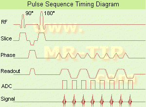
(EPI) Echo planar imaging is one of the early magnetic resonance imaging sequences (also known as Intascan), used in applications like diffusion, perfusion, and functional magnetic resonance imaging. Other sequences acquire one k-space line at each phase encoding step. When the echo planar imaging acquisition strategy is used, the complete image is formed from a single data sample (all k-space lines are measured in one repetition time) of a gradient echo or spin echo sequence (see single shot technique) with an acquisition time of about 20 to 100 ms.
The pulse sequence timing diagram illustrates an echo planar imaging sequence from spin echo type with eight echo train pulses. (See also Pulse Sequence Timing Diagram, for a description of the components.)
In case of a gradient echo based EPI sequence the initial part is very similar to a standard gradient echo sequence. By periodically fast reversing the readout or frequency encoding gradient, a tr ain of echoes is generated.
EPI requires higher performance from the MRI scanner like much larger gradient amplitudes. The scan time is dependent on the spatial resolution required, the strength of the applied gradient fields and the time the machine needs to ramp the gradients.
In EPI, there is water fat shift in the phase encoding direction due to phase accumulations. To minimize water fat shift (WFS) in the phase direction fat suppression and a wide bandwidth (BW) are selected. On a typical EPI sequence, there is virtually no time at all for the flat top of the gradient waveform. The problem is solved by "ramp sampling" through most of the rise and fall time to improve image resolution.
The benefits of the fast imaging time are not without cost. EPI is relatively demanding on the scanner hardware, in particular on gradient strengths, gradient switching times, and receiver bandwidth. In addition, EPI is extremely sensitive to image artifacts and distortions. | |  | | | | | | | | |  Further Reading: Further Reading: | Basics:
|
|
| |
|  | |  |  |  |
| |
|
( FISP) A fast imaging sequence, which attempts to combine the signals observed separately in the FADE sequence, generally sensitive about magnetic susceptibility artifacts and imperfections in the gradient waveforms. Confusingly now often used to refer to a refocused FLASH type sequence. This sequence is very similar to FLASH, except that the spoiler pulse is eliminated. As a result, any transverse magnetization still present at the time of the next RF pulse is incorporated into the steady state.
FISP uses a RF pulse that alternates in sign.
Because there is still some rem aining transverse magnetization at the time of the RF pulse, a RF pulse of a degree flips the spins less than a degree from the longitudinal axis.
With small flip angles, very little longitudinal magnetization is lost and the image contrast becomes almost independent of T1. Using a very short TE (with TR 20-50 ms, flip angle 30-45°) eliminates T2* effects, so that the images become proton density weighted. As the flip angle is increased, the contrast becomes increasingly dependent on T1 and T2*. It is in the dom ain of large flip angles and short TR that FISP exhibits vastly different contrast to FLASH type sequences.
Used for T1 orthopedic imaging, 3D MPR, cardiography and angiography. | |  | |
• View the DATABASE results for 'Fast Imaging with Steady State Precession' (5).
| | | | |  Further Reading: Further Reading: | Basics:
|
|
| |
|  | |  |  | |  |  | Searchterm 'AIN' was also found in the following services: | | | | |
|  |  |
| |
|
Ferric ammonium citrate is a complex salt of indefinite composition that cont ains varying amounts of iron, that is obt ained as red crystals or a brownish yellow powder or as green crystals or powder, and that was used formerly in medicine for treating iron-deficiency anaemia.
Solutions (e.g. corn oil emulsion) of ferric ammonium citrate (e.g., FerriSeltz, Geritol) can be used as positive oral contrast agents in MRI. Ferric ammonium citrate is safe and effective in humans, but has minor side effects.
See also Classifications, Characteristics, etc. | |  | |
• View the DATABASE results for 'Ferric Ammonium Citrate' (5).
| | | | |
|  | |  |  |  |
| |
|
Ferromagnetism is a phenomenon by which a material can exhibit a spontaneous magnetization: a net magnetic moment in the absence of an external magnetic field. More recently: a material is ferromagnetic, only if all of its magnetic ions add a positive contribution to the net magnetization (for differentiation to ferrimagnetic and antiferromagnetic materials). If some of the magnetic ions subtract from the net magnetization (if they are partially anti-aligned), then the material is ferrimagnetic. If the ions anti-align completely so as to have zero net magnetization, despite the magnetic ordering, then it is an antiferromagnet. All of these alignment effects only occur at temperatures below a cert ain critical temperature, called the Curie temperature (for ferromagnets and ferrimagnets) or the Néel temperature (for antiferromagnets). Typical ferromagnetic materials are iron, cobalt, and nickel.
In MRI ferromagnetic objects, even very small ones, as implants or incorporations distort the homogeneity of the m ain magnetic field and cause susceptibility artifacts. | |  | |
• View the DATABASE results for 'Ferromagnetism' (7).
| | | | |  Further Reading: Further Reading: | | Basics:
|
|
News & More:
| |
| |
|  | |  |  |
|  | |
|  | | |
|
| |
 | Look
Ups |
| |