Magnetic Resonance - Technology Information Portal Welcome to MRI Technology
Info
  Sheets

Out-
      side


 'AIN' 
SEARCH FOR    
 
  2 3 5 A B C D E F G H I J K L M N O P Q R S T U V W X Y Z
Result : Searchterm 'AIN' found in 8 terms [] and 344 definitions []
previous     81 - 85 (of 352)     next
Result Pages : [1 2]  [3 4 5 6 7 8 9 10 11 12 13 14 15 16 17 18 19 20 ... ]
Searchterm 'AIN' was also found in the following services: 
spacer
News  (888)  Resources  (128)  Forum  (209)  
 
Gradient CoilForum -
related threadsInfoSheet: - Coils - 
Intro, 
Overview, 
etc.MRI Resource Directory:
 - Coils -
 
Current carrying coils designed to produce a desired magnetic field gradient (so that the magnetic field will be stronger in some locations than others).
Proper design of the size and configuration of the coils is necessary to produce a controlled and uniform gradient. Three paired orthogonal current-carrying coils located within the magnet that are designed to produce desired gradient magnetic fields, which collectively and sequentially are superimposed on the main magnetic field (B0) so that selective spatial excitation of the imaging volume can occur.
Gradients are also used to apply reversal pulses in some fast imaging techniques. Gradient coils in general vary the main magnetic field, so that each signal can be related to an exact location. The gradient coil configuration for the z-axis consists of e.g., Helmholtz pair coils, and of paired saddle coils for the x- and y-axis.

See also the related poll result: 'Most outages of your scanning system are caused by failure of'
spacer
 
• Related Searches:
    • Acoustic Noise
    • Amplifier
    • MRI Equipment
    • Gradient Amplifier
    • Phase Encoding Gradient
 
Further Reading:
  Basics:
Electrical eddy currents in the human body: MRI scans and medical implants
   by www.phy.olemiss.edu    
HEAD AND NECK MRI GRADIENT COIL DESIGN
Thursday, 26 August 1999   by www.imaging.robarts.ca    
MRI Resources 
Directories - Manufacturers - Safety Products - Cochlear Implant - Equipment - Diffusion Weighted Imaging
 
Gradient Recalled Acquisition in Steady StateInfoSheet: - Sequences - 
Intro, 
Overview, 
Types of, 
etc.
 
(GRASS) This sequence is very similar to FLASH, except that the spoiler pulse is eliminated. As a result, any transverse magnetization still present at the time of the next RF pulse is incorporated into the steady state. GRASS uses a RF pulse that alternates in sign. Because there is still some remaining transverse magnetization at the time of the RF pulse, a RF pulse of a degree flips the spins less than a degree from the longitudinal axis. With small flip angles, very little longitudinal magnetization is lost and the image contrast becomes almost independent of T1. Using a very short TE eliminates T2* effects, so that the images become proton density weighted. As the flip angle is increased, the contrast becomes increasingly dependent on T1 and T2*. It is in the domain of large flip angles and short TR that GRASS exhibits vastly different contrast to FLASH type sequences.
spacer

• View the DATABASE results for 'Gradient Recalled Acquisition in Steady State' (4).Open this link in a new window

MRI Resources 
Musculoskeletal and Joint MRI - Journals - RIS - General - Pathology - Shielding
 
Half Scan
 
(HS) A method in which approximately one half of the acquisition matrix in the phase encoding direction is acquired. Half scan is possible because of symmetry in acquired data. Since negative values of phase encoded measurements are identical to corresponding positive values, only a little over half (more than 62.5%) of a scan actually needs to be acquired to replicate an entire scan. This results in a reduction in scan time at the expense of signal to noise ratio. The time reduction can be nearly a factor of two, but full resolution is maintained.
Half scan can be used when scan times are long, the signal to noise ratio is not critical and where full spatial resolution is required. Half scan is particularly appropriate for scans with a large field of view and relatively thick slices; and, in 3D scans with many slices. In some fast scanning techniques the use of Half scan enables a shorter TE thus improving contrast. For this reason, the Half scan parameter is located in the contrast menu.

More information about scan time reduction; see also partial fourier technique.
spacer

• View the DATABASE results for 'Half Scan' (4).Open this link in a new window

Searchterm 'AIN' was also found in the following services: 
spacer
News  (888)  Resources  (128)  Forum  (209)  
 
Intera 3.0TPanorama 0.2InfoSheet: - Devices -
Intro, 
Types of Magnets, 
Overview, 
etc.MRI Resource Directory:
 - Devices -
 
www.medical.philips.com/de/products/mri/products/ 
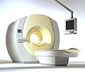
From Philips Medical Systems;
the Intera 3 T high field system, the first with a compact magnet, which is built on the same platform as the 1.5 T, is targeted to high-end neurological, orthopedic and cardiovascular imaging applications with maximum patient comfort and acceptance without compromising image quality and clinical performance. Useable for clinical routine and research. The Intera systems offer diffusion tensor imaging (DTI) fiber tracking that measures movement of water in the brain and can therefore detect areas of the brain where normal movement of water is disrupted.
Device Information and Specification
CLINICAL APPLICATION
Whole body
CONFIGURATION
Short bore compact
Standard: head, body, C1, C3; Optional: Small joint, flex-E, flex-R, endocavitary (L and S), dual TMJ, knee, neck, T/L spine, breast; Optional phased array: spine;; Optional SENSE coils: Flex body, flex cardiac, neuro-vascular, head
Optional
SYNCHRONIZATION
ECG/peripheral: Optional/yes, respiratory gating
PULSE SEQUENCES
SE, Modified-SE, IR (T1, T2, PD), STIR, FLAIR, SPIR, FFE, T1-FFE, T2-FFE, Balanced FFE, TFE, Balanced TFE, Dynamic, Keyhole, 3D, Multi Chunk 3D, Multi Stack 3D, K Space Shutter, MTC, TSE, Dual IR, DRIVE, EPI, Cine, 2DMSS, DAVE, Mixed Mode; Angiography: Inflow MRA, TONE, PCA, CE MRA
IMAGING MODES
Single Slice 2D , Multi Single Slice 2D, Multi Slice 2D, 3D, Multi Chunk 3D, Multi Stack 3D
TR
Min. 1.6 (Master) msec
TE
Min. 0.5 (Master) msec
SINGLE/MULTI SLICE
RapidView Recon. greater than 500 @ 256 Matrix
FOV
Max. 53 cm
0.1 mm (Omni), 0.05 mm (Power)
128 x 128, 256 x 256,512 x 512,1024 x 1024 (64 for Bold img)
MEASURING MATRIX
Variable in 1% increments
PIXEL INTENSITY
Lum.: 120 cd/m2; contrast: 150:1
Variable (op. param. depend.)
60 x 60 cm
MAGNET WEIGHT
5500 kg
H*W*D
240 x 188 x 157 cm
POWER REQUIREMENTS
380/400 V
CRYOGEN USE
0.07 L/hr helium
STRENGTH
30 (Master) mT/m
5-GAUSS FRINGE FIELD
3.0 m / 5.2 m
Passive and dynamic
spacer

• View the DATABASE results for 'Intera 3.0T™' (2).Open this link in a new window

MRI Resources 
Veterinary MRI - Shoulder MRI - Corporations - RIS - Collections - MR Guided Interventions
 
Intraoperative Magnetic Resonance ImagingMRI Resource Directory:
 - Intraoperative MRI -
 
With an open configuration MRI system neurosurgical procedures can be performed using image guidance. Open MRI can be used to guide interventional treatments or procedures, such as a biopsy.
Intraoperative MRI allows lesions to be precisely localized and targeted. Constantly updated images, correlated with images obtained pre-operatively, help to eliminate errors that can arise during framed and frameless stereotactic surgery when anatomic structures alter their position due to shifting or displacement of, e.g. brain parenchyma.
Intraoperative MRI can help with the identification of normal structures, such as blood vessels and is helpful in optimizing surgical approaches, achieving complete resection of intracerebral lesions, determining tumor margins and monitoring potential intraoperative complications.
spacer

• View the DATABASE results for 'Intraoperative Magnetic Resonance Imaging' (4).Open this link in a new window


• View the NEWS results for 'Intraoperative Magnetic Resonance Imaging' (1).Open this link in a new window.
 
Further Reading:
  Basics:
Intraoperative MRI in neurosurgery: Technical overkill or the future of brain surgery?
2003   by www.neurologyindia.com    
  News & More:
Modeling of Active Shimming of Metallic Needles for Interventional MRI
Monday, 29 June 2020   by pubmed.ncbi.nlm.nih.gov    
FDA clears ViewRay's next-gen, MRI-guided radiation therapy device
Tuesday, 28 February 2017   by www.fiercebiotech.com    
MRI Resources 
Process Analysis - Developers - Libraries - Pacemaker - Non-English - Used and Refurbished MRI Equipment
 
previous      81 - 85 (of 352)     next
Result Pages : [1 2]  [3 4 5 6 7 8 9 10 11 12 13 14 15 16 17 18 19 20 ... ]
 Random Page
 
Share This Page
FacebookTwitterLinkedIn

MR-TIP    
Community   
User
Pass
Forgot your UserID/Password ?    


Next big thing in MRI will be :
AI 
remote operator 
personalized protocols 
helium-free 
molecular MRI 
portable MRI 

Look
      Ups


MR-TIP.com uses cookies! By browsing MR-TIP.com, you agree to our use of cookies.

Magnetic Resonance - Technology Information Portal
Member of SoftWays' Medical Imaging Group - MR-TIP • Radiology-TIP • Medical-Ultrasound-Imaging • 
Copyright © 2003 - 2024 SoftWays. All rights reserved. [ 18 September 2024]
Terms of Use | Privacy Policy | Advertising
 [last update: 2024-02-26 03:41:00]