 | Info
Sheets |
| | | | | | | | | | | | | | | | | | | | | | | | |
 | Out-
side |
| | | | |
|
| | | | | |  | Searchterm 'Arc' was also found in the following services: | | | | |
|  |  |
| |
|

From GE Healthcare;
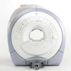
The GE Signa HDx MRI system is a whole body magnetic resonance scanner designed to support high resolution, high signal to noise ratio, and short scan times.
The 1.5T Signa HDx MR Systems is a modification of the currently marketed GE 1.5T machines, with the main difference being the change to the receive chain architecture that includes a thirty two independent receive channels, and allows for future expansion in 16 channel increments. The overall system has been improved with a simplified user interface
and a single 23" liquid crystal display, improved multi channel surface coil connectivity, and an improved image reconstruction architecture known as the Volume Recon Engine (VRE).
Device Information and Specification CLINICAL APPLICATION Whole body CONFIGURATION Compact short bore Standard: SE, IR, 2D/3D GRE and SPGR, Angiography: 2D/3D TOF, 2D/3D Phase Contrast; 2D/3D FSE, 2D/3D FGRE and FSPGR, SSFP, FLAIR, EPI, optional: 2D/3D Fiesta, FGRET, Spiral, Tensor, 2D 0.7 mm to 20 mm; 3D 0.1 mm to 5 mm 128x512 steps 32 phase encode POWER REQUIREMENTS 480 or 380/415 less than 0.03 L/hr liquid helium | |  | | | |
|  |  | Searchterm 'Arc' was also found in the following services: | | | | |
|  |  |
| |
|
 [This entry is marked for removal.]
Toshiba Medical Systems started 1914 and was a leading diagnostic imaging manufacturer with departments including rese arch, development, production, service and support of medical imaging equipment and systems. 1976 Toshiba America Medical Systems, Inc. (TAMS) was founded to coordinate sales and service for previously established business operations in the USA.
In M arch 2016 Toshiba sold its Toshiba Medical Systems for $6 billion to Japan's Canon. After regulatory approval the company got renamed to Canon Medical Systems.
MRI Scanners:
0.2T to 1.0T:
1.5T:
3.0T:
| |  | |
• View the DATABASE results for 'Toshiba America Medical Systems Inc.' (8).
| | |
• View the NEWS results for 'Toshiba America Medical Systems Inc.' (10).
| | | | |  Further Reading: Further Reading: | Basics:
|
|
| |
|  | |  |  |  |
| |
|

California-based rese arch and development company. Alliance has several patents regarding ' microbubble' compositions such as Imagent® that are intended to enhance ultrasound imaging and Imagent GI as a gastrointestinal MRI contrast agent. Another product is Oxygent™, a synthetic 'blood substitute'.
MRI Contrast Agents:
Contact Information
MAIL
Alliance Pharmaceutical Corp.
6175 Lusk Blvd.
San Diego, CA 92121
USA
| |  | |
• View the DATABASE results for 'Alliance Pharmaceutical Corp.' (2).
| | | | |  Further Reading: Further Reading: | Basics:
|
|
| |
|  |  | Searchterm 'Arc' was also found in the following services: | | | | |
|  |  |
| |
|

The company is a member of the Bracco Group, a highly innovative healthcare group and world leader in global integrated solutions for the diagnostic medical imaging field. The Bracco Group is headquartered in Milan, Italy. Its North American operations consist of Bracco Diagnostics and Bracco Rese arch USA, both located in Princeton, New Jersey. Bracco Diagnostics is one of the fastest growing developers and marketers of diagnostic pharmaceuticals in North America, with products for various imaging applications, including Isovue® (iopamidol - X-ray contrast agent), ProHance® ( gadoteridol - MRI contrast agent), SonoVue® ( ultrasound contrast agent) and nuclear medicine products.
Gadoteridol has been available in Europe and the USA for several years.
Holder of the Marketing Authorization:
Bracco International B.V. - Strawinskylaan 3051 - 1077 ZX Amsterdam
The Netherlands. (Contact: Kirk Deeter, Phone: +NL-303-838-8708)
MRI Contrast Agents:
Contact Information
Please see Bracco Diagnostics, Inc.'s
| |  | |
• View the DATABASE results for 'Bracco Diagnostics, Inc.' (2).
| | | | |  Further Reading: Further Reading: | News & More:
|
|
| |
|  |  | Searchterm 'Arc' was also found in the following services: | | | | |
|  |  |
| |
|
In the last years, cardiac MRI techniques have progressively improved. No other noninvasive imaging modality provides the same degree of contrast and temporal resolution for the assessment of cardiovascular anatomy and pathology. Contraindications MRI are the same as for other magnetic resonance techniques.
The primary advantage of MRI is extremely high contrast resolution between different tissue types, including blood. Moreover, MRI is a true 3 dimensional imaging modality and images can be obtained in any oblique plane along the true cardiac axes while preserving high temporal and spatial resolution with precise demonstration of cardiac anatomy without the administration of contrast media.
Due to these properties, MRI can precisely characterize cardiac function and quantify cavity volumes, ejection fraction, and left ventricular mass. In addition, cardiac MRI has the ability to quantify flow (see flow quantification), including bulk flow in vessels, pressure gradients across stenosis, regurgitant fractions and shunt fractions. Valve morphology and area can be determined and the severity of stenosis quantified. In certain disease states, such as myocardial inf arction, the contrast resolution of MRI is further improved by the addition of extrinsic contrast agents (see myocardial late enhancement).
A dedicated cardiac coil, and a field strength higher than 1 Tesla is recommended to have sufficient signal. Cardiac MRI acquires ECG gating. Cardiac gating (ECGs) obtained within the MRI scanner, can be degraded by the superimposed electrical potential of flowing blood in the magnetic field. Therefore, excellent contact between the skin and ECG leads is necessary. For male patients, the skin at the lead sites can be shaved. A good cooperation of the patient is necessary because breath holding at the end of expiration is practiced during the most sequences.
See also Displacement Encoding with Stimulated Echoes.
For Ultrasound Imaging (USI) see Cardiac Ultrasound at Medical-Ultrasound-Imaging.com.
See also the related poll results: ' In 2010 your scanner will probably work with a field strength of' and ' MRI will have replaced 50% of x-ray exams by' | | | |  | |
• View the DATABASE results for 'Cardiac MRI' (15).
| | |
• View the NEWS results for 'Cardiac MRI' (15).
| | | | |  Further Reading: Further Reading: | | Basics:
|
|
News & More:
|  |
MRI technology visualizes heart metabolism in real time
Friday, 18 November 2022 by medicalxpress.com |  |  |
Even early forms of liver disease affect heart health, Cedars-Sinai study finds
Thursday, 8 December 2022 by www.eurekalert.org |  |  |
MRI sheds light on COVID vaccine-associated heart muscle injury
Tuesday, 15 February 2022 by www.sciencedaily.com |  |  |
Radiologists must master cardiac CT, MRI to keep pace with demand: The heart is not a magical organ
Monday, 1 March 2021 by www.radiologybusiness.com |  |  |
Diffusion weighted imaging (DWI) and diffusion tensor imaging (DTI) in the heart (myocardium)
Sunday, 30 August 2020 by github.com |  |  |
Non-invasive diagnostic procedures for suspected CHD: Search reveals informative evidence
Wednesday, 8 July 2020 by medicalxpress.co |  |  |
Cardiac MRI Becoming More Widely Available Thanks to AI and Reduced Exam Times
Wednesday, 19 February 2020 by www.dicardiology.com |  |  |
Controlling patient's breathing makes cardiac MRI more accurate
Friday, 13 May 2016 by www.upi.com |  |  |
Precise visualization of myocardial injury: World's first patient-based cardiac MRI study using 7T MRI
Wednesday, 10 February 2016 by medicalxpress.com |  |  |
New technique could allow for safer, more accurate heart scans
Thursday, 10 December 2015 by www.gizmag.com |
|
| |
|  | |  |  |
|  | |
|  | | |
|
| |
 | Look
Ups |
| |