 | Info
Sheets |
| | | | | | | | | | | | | | | | | | | | | | | | |
 | Out-
side |
| | | | |
|
| | | | | |  | Searchterm 'Bo' was also found in the following services: | | | | |
|  |  |
| |
|
From Philips Medical Systems;
The clinical capabilities of MR will further expand. Inside and out, the Achieva is a friendly, open system designed for optimal patient comfort and maximized workflow with high functionality.
The Achieva 1.5T can be upgraded to Achieva I/T, with three configurations optimized for MR guided interventions and therapy:
•
Achieva I/T Neurosurgery
•
Achieva I/T Cardiovascular (or XMR - combining an Achieva 1.5T CV system and an X-Ray system)
Device Information and Specification
CLINICAL APPLICATION
Whole body
CONFIGURATION
Short bore compact
Standard: Head, body, C1, C3; Optional: Small joint, flex-E, flex-R, endocavitary (L and S), dual TMJ, knee, neck, T/L spine, breast; optional phased array: Spine, pediatric, 3rd party connector; Optional SENSEâ„¢ coils for all applications
SE, Modified-SE, IR (T1, T2, PD), STIR, FLAIR, SPIR, FFE, T1-FFE, T2-FFE, Balanced FFE, TFE, Balanced TFE, Dynamic, Keyhole, 3D, Multi Chunk 3D, Multi Stack 3D, K Space Shutter, MTC, TSE, Dual IR, DRIVE, EPI, Cine, 2DMSS, DAVE, Mixed Mode; Angiography: Inflow MRA, TONE, PCA, CE MRA
128 x 128, 256 x 256,512 x 512,1024 x 1024 (64 for Bold img)
Variable in 1% increments
Lum.: 120 cd/m2; contrast: 150:1
Variable (op. param. depend.)
POWER REQUIREMENTS
380/400 V
| | | | | |
| | | | | |
| |
|
Device Information and Specification
CLINICAL APPLICATION
Whole body
CONFIGURATION
Short bore compact
Standard: Head, body, cardiac, optional phased array: Spine, pediatric, 3rd party connector; Optional SENSE? coils for all applications
SE, Modified-SE, IR (T1, T2, PD), STIR, FLAIR, SPIR, FFE, T1-FFE, T2-FFE, Balanced FFE, TFE, Balanced TFE, Dynamic, Keyhole, 3D, Multi Chunk 3D, Multi Stack 3D, K Space Shutter, MTC, TSE, Dual IR, DRIVE, EPI, Cine, 2DMSS, DAVE, Mixed Mode; Angiography: Inflow MRA, TONE, PCA, CE MRA
128 x 128, 256 x 256,512 x 512,1024 x 1024 (64 for Bold img)
Variable in 1% increments
Lum.: 120 cd/m2; contrast: 150:1
Variable (op. param. depend.)
POWER REQUIREMENTS
380/400 V
| | | |
• View the DATABASE results for 'Intera Achieva CV™' (2).
| | | | |  Further Reading: Further Reading: | News & More:
|
|
| |
| | | | | |
| |
|

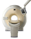
From Siemens Medical Systems;
MAGNETOM Avanto with Tim - Total imaging matrix technology.
For true whole- body anatomical coverage. For ultra-fast image
acquisition. Aids the physician in fast and precise
evaluation of systemic diseases like colon cancer, metastasis imaging, vessel diseases, and preventional exams. For claustrophobic patients,
MAGNETOM Avanto enables feetfirst exams for nearly all MR procedures. For obese patients, MAGNETOM Avanto supports up to 200 kg (400 lbs), without table movement restrictions. The AudioComfort technology enables up to a 30 dB(A) acoustic noise reduction, that means nearly all clinical routine sequences are running under 99 dB(A).
Device Information and Specification
CLINICAL APPLICATION
Whole body
CONFIGURATION
Compact short bore
Body, Tim [32 x 8], Tim [76 x18], Tim [76 coil elements with up to 32 RF channels]
GRE, IR, FIR, STIR, TrueIR/FISP, FSE, FLAIR, MT, SS-FSE, MT-SE, MTC, MSE, EPI, 3D DESS//CISS/PSIF, GMR
IMAGING MODES
Single, multislice, volume study, multi angle, multi oblique
1024 x 1024 full screen display
POWER REQUIREMENTS
380/400/420/440/480 V
Passive, act.; 1st order std./2nd opt.
| | | |
• View the DATABASE results for 'MAGNETOM Avanto™' (2).
| | | | | | Further Reading: | | Basics:
|
|
News & More:
| |
| |
| | | Searchterm 'Bo' was also found in the following services: | | | | |
| | |
| |
|

It is important to remember when working around a superconducting magnet that the magnetic field is always on. Under usual working conditions the field is never turned off. Attention must be paid to keep all ferromagnetic items at an adequate distance from the magnet. Ferromagnetic objects which came accidentally under the influence of these strong magnets can injure or kill individuals in or nearby the magnet, or can seriously damage every hardware, the magnet itself, the cooling system, etc..
See MRI resources Accidents.
The doors leading to a magnet room should be closed at all times except when entering or exiting the room. Every person working in or entering the magnet room or adjacent rooms with a magnetic field has to be instructed a bout the dangers. This should include the patient, intensive-care staff, and maintenance-, service- and cleaning personnel, etc..
The 5 Gauss limit defines the 'safe' level of static magnetic field exposure. The value of the absorbed dose is fixed by the authorities to avoid heating of the patient's tissue and is defined by the specific absorption rate.
Leads or wires that are used in the magnet bore during imaging procedures, should not form large-radius wire loops. Leg-to-leg and leg-to-arm skin contact should be prevented in order to avoid the risk of burning due to the generation of high current loops if the legs or arms are allowed to touch. The patient's skin should not be in contact with the inner bore of the magnet.
The outflow from cryogens like liquid helium is improbable during normal operation and not a real danger for patients.
The safety of MRI contrast agents is tested in drug trials and they have a high compatibility with very few side effects. The variations of the side effects and possible contraindications are similar to X-ray contrast medium, but very rare. In general, an adverse reaction increases with the quantity of the MRI contrast medium and also with the osmolarity of the compound.
See also 5 Gauss Fringe Field, 5 Gauss Line, Cardiac Risks, Cardiac Stent, dB/dt, Legal Requirements, Low Field MRI, Magnetohydrodynamic Effect, MR Compatibility, MR Guided Interventions, Claustrophobia, MRI Risks and Shielding. | | | | | | | | |
• View the DATABASE results for 'MRI Safety' (42).
| | |
• View the NEWS results for 'MRI Safety' (13).
| | | | | | Further Reading: | | Basics:
|
|
News & More:
| |
| |
| | | | | |
| |
|
The definition of a scan is to form an image or an electronic representation. The MRI scan uses magnetic resonance principles to produce extremely detailed pictures of the body tissue without the need for X-ray exposure or other damaging forms of radiation.
MRI scans show structures of the different tissues in the body. The tissue that has the least hydrogen atoms (e.g., bones) appears dark, while the tissue with many hydrogen atoms (e.g., fat) looks bright. The MRI pictures of the brain show details and abnormal structures ( brain MRI), for example, tumors, multiple sclerosis lesions, bleedings, or brain tissue that has suffered lack of oxygen after a stroke.
A cardiac MRI scan demonstrates the heart as well as blood vessels ( cardiovascular imaging) and is used to detect heart defects with e.g., changes in the thickness and infarctions of the muscles around the heart. With MRI scans, nearly all kind of body parts can be tested, for example the joints like knee and shoulder, lumbar, thoracic and cervical spine, the pelvis including fetal MRI, and the soft parts of the body such as the liver, kidneys, and spleen.
The MRI procedure includes three to nine imaging sequences and may take up to one hour. See also Lumbar Spine MRI, MRI Safety and Open MRI. | | | | | | | | | | |
• View the DATABASE results for 'MRI Scan' (31).
| | |
• View the NEWS results for 'MRI Scan' (95).
| | | | | | Further Reading: | Basics:
|
|
News & More:
|  |
A Knee MRI in Half the Time? It's Possible
Thursday, 8 April 2021 by www.diagnosticimaging.com | | |
Michigan radiologist warns about 'incidental findings' in full body MRI scans
Wednesday, 4 October 2023 by www.wilx.com | | |
ACCELERATING MRI SCANS WITH ARTIFICIAL INTELLIGENCE
Friday, 28 August 2020 by www.analyticsinsight.net | | |
Radiographer's Lego Open MRI Product Idea Reaches New Milestone
Monday, 11 November 2019 by www.itnonline.com | | |
Why we need erasable MRI scans
Wednesday, 25 April 2018 by phys.org | | |
MRI as accurate as CT for Crohn's disease detection, management
Tuesday, 6 June 2017 by www.healthimaging.com | | |
MRI scans predict patients' ability to fight the spread of cancer
Tuesday, 12 December 2017 by eurekalert.org | | |
Audio/Video System helps patients relax during MRI scans
Monday, 8 December 2014 by news.thomasnet.com | | |
MRI scans could be a 'game-changer' in prostate cancer testing
Tuesday, 5 August 2014 by www.abc.net.au | | |
7-Tesla MRI scanner allows even more accurate diagnosis of breast cancer
Thursday, 6 March 2014 by www.healthcanal.com |
|
| |
| | | | |
| | | |
|
| |
 | Look
Ups |
| |


