 | Info
Sheets |
| | | | | | | | | | | | | | | | | | | | | | | | |
 | Out-
side |
| | | | |
|
| | | | | |  | Searchterm 'Bo' was also found in the following services: | | | | |
|  |  |
| |
|
The definition of imaging is the visual representation of an object. Medical imaging began after the discovery of x-rays by Konrad Roentgen 1896. The first fifty years of radiological imaging, pictures have been created by focusing x-rays on the examined body part and direct depiction onto a single piece of film inside a special cassette. The next development involved the use of fluorescent screens and special glasses to see x-ray images in real time.
A major development was the application of contrast agents for a better image contrast and organ visualization. In the 1950s, first nuclear medicine studies showed the up-take of very low-level radioactive chemicals in organs, using special gamma cameras. This medical imaging technology allows information of biologic processes in vivo. Today, PET and SPECT play an important role in both clinical research and diagnosis of biochemical and physiologic processes. In 1955, the first x-ray image intensifier allowed the pick up and display of x-ray movies.
In the 1960s, the principals of sonar were applied to diagnostic imaging. Ultrasonic waves generated by a quartz crystal are reflected at the interfaces between different tissues, received by the ultrasound machine, and turned into pictures with the use of computers and reconstruction software. Ultrasound imaging is an important diagnostic tool, and there are great opportunities for its further development. Looking into the
future, the grand challenges include targeted contrast agents, real-time 3D ultrasound imaging, and molecular imaging.
Digital imaging techniques were implemented in the 1970s into conventional fluoroscopic image intensifier and by Godfrey Hounsfield with the first computed tomography. Digital images are electronic snapshots sampled and mapped as a grid of dots or pixels. The introduction of x-ray CT revolutionised medical imaging with cross sectional images of the human body and high contrast between different types of soft tissue. These developments were made possible by analog to digital converters and computers. The multislice spiral CT technology has expands the clinical applications dramatically.
The first MRI devices were tested on clinical patients in 1980. The spread of CT machines is the spur to the rapid development of MRI imaging and the introduction of tomographic imaging techniques into diagnostic nuclear medicine. With technological improvements including higher field strength, more open MRI magnets, faster gradient systems, and novel data-acquisition techniques, MRI is a real-time interactive imaging modality that provides both detailed structural and functional information of the body.
Today, imaging in medicine has advanced to a stage that was inconceivable 100 years ago, with growing medical imaging modalities:
•
Single photon emission computed tomography (SPECT)
•
Positron emission tomography (PET)
All this type of scans are an integral part of modern healthcare.
Because of the rapid development of digital imaging modalities, the increasing need for an efficient management leads to the widening of radiology information systems (RIS) and archival of images in digital form in picture archiving and communication systems (PACS).
In telemedicine, healthcare professionals are linked over a computer network. Using cutting-edge computing and communications technologies, in videoconferences, where audio and visual images are transmitted in real time, medical images of MRI scans, x-ray examinations, CT scans and other pictures are shareable.
See also Hybrid Imaging.
See also the related poll results: ' In 2010 your scanner will probably work with a field strength of', ' MRI will have replaced 50% of x-ray exams by' | | | | | | | | | | | | | | | |  Further Reading: Further Reading: | | Basics:
|
|
News & More:
| |
| |
| | | | | |
| |
|
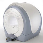
Open MRI scanners have been developed for people who are anxious or obese or for examination of small parts of the body, such as the extremities ( knee, shoulder). In addition, some systems offer imaging in different positions and sequences of movements.
The basic technology of an open MRI machine is similar to that of a traditional MRI device.
The major difference for the patient is that instead of lying in a narrow tunnel, the imaging table has more space around the body so that the magnet does not completely surround the person being tested.
Types of constructions:
•
Semi open high field MRI scanners provide an ultra short bore (tunnel) and widely flared ends. In this type of MRI systems, patients lie with the head in the space outside the bore, if for example the hips are examined.
•
Open low field MRI machines have often a wide open design, e.g. an open C-arm scanner is shaped like two large discs separated by a large pillar. Patients have an open sided feeling and more space around them allows a wider range of positions.
•
Advanced open MRI scanners combine the advantages of both, the high field strength, newest gradient technology and wide open design. Even scans of patients in upright, weight-bearing positions are possible (e.g. Upright™ MRI formerly Stand-Up MRI).
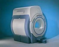
Difficulties with a traditional MRI scan include claustrophobia and patient size or, for health related reasons, patients who are not able to receive this type of diagnostic test. The MRI unit is a limited space, and some patients may be too large to fit in a narrow tunnel. In addition, weight limits can restrict the use of some scanners. The open MRI magnet has become the best option for those patients.
All of the highest resolution MRI scanners are tunnels and tend to accentuate the claustrophobic reaction. While patients may find the open MRI scanners easier to tolerate, some machines use a lower field magnet and generates lower image quality or have longer scan time. The better performance of an advanced open MRI scanner allows good image quality caused by the higher signal to noise ratio with maximum patient comfort.
See also Claustrophobia, MRI scan and Knee MRI. | | | |
• View the DATABASE results for 'Open MRI' (37).
| | |
• View the NEWS results for 'Open MRI' (16).
| | | | | | Further Reading: | Basics:
|
|
News & More:
| |
| |
| | | | | |
| |
|
If available, some graphic aids can be helpful to show image orientations.
1) A graphic icon of the labeled primary axes (A, L, H) with relative lengths given by direction sines and orientation as if viewed from the normal to the image plane can help orient the viewer, both to identify image plane orientation and to indicate possible in plane rotation.
2) Ingraphic prescription of obliques from other images, a sample original image with an overlaid line or set of lines indicating the intersection of the original and oblique image planes can help orient the viewer.
•
The basic anatomical directions are:
right(R) to left (L), posterior (P) to anterior (A), and feet (F) to head (H).
•
A standard display orientation for images in the basic slice orientation is:
1) transverse: A to top of image and L to right,
2) coronal: H to top of image and L to right and
3) sagittal: H to top of image and A to left.
The location in the R/L and P/A directions can be specified relative to the axis of the magnet.
The F/H location can be specified relative to a convenient patient structure.
The orientation of single oblique slices can be specified by rotating a slice in one of the basic orientations toward one of the other two basic orthogonal planes a bout an axis defined by the intersection of the 2 planes.
Double oblique slices can be specified as the result of tipping a single oblique plane toward the remaining basic orientation plane, a bout an axis defined by the intersection of the oblique plane and the remaining basic plane. In double oblique angulations, the first rotation is chosen a bout the vertical image axis and the second a bout the (new) horizontal axis.
Angles are chosen to have magnitudes less than 90° (for single oblique slices less than 45°); the sign of the angle is taken to be positive when the rotation brings positive axes closer together. | | | | | |
• View the DATABASE results for 'Orientation' (16).
| | | | |
| | | Searchterm 'Bo' was also found in the following services: | | | | |
| | |
| |
|
From GE Healthcare;
The Signa HDx MRI system is GE's leading edge whole body magnetic resonance scanner designed to support high resolution, high signal to noise ratio, and short scan times.
Signa HDx 3.0T offers new technologies like ultra-fast image reconstruction through the new XVRE recon engine, advancements in parallel imaging algorithms and the broadest range of premium applications. The HD applications, PROPELLER (high-quality brain imaging extremely resistant to motion artifacts), TRICKS (contrast-enhanced angiographic vascular lower leg imaging), VIBRANT (for breast MRI), LAVA (high resolution liver imaging with shorter breath holds and better organ coverage) and MR Echo (high-definition cardiac images in real time) offer unique capabilities.
Device Information and Specification CLINICAL APPLICATION Whole body
CONFIGURATION Compact short bore SE, IR, 2D/3D GRE, RF-spoiled GRE, 2DFGRE, 2DFSPGR, 3DFGRE, 3DFSPGR, 3DTOFGRE, 3DFSPGR, 2DFSE, 2DFSE-XL, 2DFSE-IR, T1-FLAIR, SSFSE, EPI, DW-EPI, BRAVO, Angiography: 2D/3D TOF, 2D/3D phase contrast vascular IMAGING MODES Single, multislice, volume study, fast scan, multi slab, cine, localizer H*W*D 240 x 2216,6 x 201,6 cm POWER REQUIREMENTS 480 or 380/415, 3 phase ||
COOLING SYSTEM TYPE Closed-loop water-cooled grad. | | | | | |
| | | | | |
| |
|
From GE Healthcare;
'EXCITE technology has the potential to open the door to new imaging techniques and clinical applications, leaping beyond conventional two and three-dimensional MRI to true 4D imaging that will improve the diagnosis of disease throughout the human body from head to foot.' Robert R. Edelman, M.D., Professor of Radiology at Northwestern University Medical School and Chairman, Department of Radiology, at Evanston Northwestern Healthcare.
Device Information and Specification CLINICAL APPLICATION Whole body Head and body coil standard; all other coils optional; open architecture makes system compatible with a wide selection of coils Optional 2D/3D brain and prostate Standard: SE, IR, 2D/3D GRE and SPGR, Angiography: 2D/3D TOF, 2D/3D Phase Contrast;; 2D/3D FSE, 2D/3D FGRE and FSPGR, SSFP, FLAIR, EPI, optional: 2D/3D Fiesta, FGRET, Spiral, TensorTR 1.3 to 12000 msec in increments of 1 msec TE 0.4 to 2000 msec in increments of 1 msec 2D 0.7 mm to 20 mm; 3D 0.1 mm to 5 mm 128x512 steps 32 phase encode 0.08 mm; 0.02 mm optional POWER REQUIREMENTS 480 or 380/415 less than 0.03 L/hr liquid heliumSTRENGTH SmartSpeed 23 mT/m, HiSpeed Plus 33 mT/m, EchoSpeed Plus 33 mT/m 4.0 m x 2.8 m axial x radial | | | |
• View the DATABASE results for 'Signa Infinity 1.5T™ with Excite' (2).
| | | | |
| | | | |
| | | |
|
| |
 | Look
Ups |
| |


