 | Info
Sheets |
| | | | | | | | | | | | | | | | | | | | | | | | |
 | Out-
side |
| | | | |
|
| | | | | |  | Searchterm 'Bo' was also found in the following services: | | | | |
|  |  |
| |
|
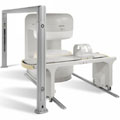
Device Information and Specification CLINICAL APPLICATION Whole body GRE, IR, FIR, STIR, TrueIR/FISP, FSE, FLAIR, MT, SS-FSE, MT-SE, MTC, MSE, EPI, GMR, fat/water sat./exc. IMAGING MODES Single, multislice, volume study, multi angle, multi obliqueTR 2.4 msec std.; 2.0 opt.; 1.8 w/30 mT/m at 256matrix TE 1.1 msec std.; 0.9 opt.; 0.78 w/30 mT/m at 256matrix 178 images/sec at 256 x 256 at 100% FOV1024 x 1024 full screen display 21 micrometer in plane, 11 micrometer optional 4050kg, 5500kg in operation H*W*D 236 x 215 x 160 cm w/covers POWER REQUIREMENTS 380/400/420/440/480 V STRENGTH 20/35 mT/m standard, 30/52 opt. Passive, act.; 1st order std./2nd opt. | | | | | | | | | | |  Further Reading: Further Reading: | Basics:
|
|
| |
| | | | | |
| |
|
( MRI) Magnetic resonance imaging is a noninvasive medical imaging technique that uses the interaction between radio frequency pulses, a strong magnetic field and body tissue to obtain images of slices/planes from inside the body. These magnets generate fields from approx. 2000 times up to 30000 times stronger than that of the Earth. The use of nuclear magnetic resonance principles produces extremely detailed pictures of the body tissue without the need for x-ray exposure and gives diagnostic information of various organs.
Measured are mobile hydrogen nuclei (protons are the hydrogen atoms of water, the 'H' in H 20), the majority of elements in the body. Only a small part of them contribute to the measured signal, caused by their different alignment in the magnetic field. Protons are capable of absorbing energy if exposed to short radio wave pulses (electromagnetic energy) at their resonance frequency. After the absorption of this energy, the nuclei release this energy so that they return to their initial state of equilibrium.
This transmission of energy by the nuclei as they return to their initial state is what is observed as the MRI signal. The subtle differing characteristic of that signal from different tissues combined with complex mathematical formulas analyzed on modern computers is what enables MRI imaging to distinguish between various organs. Any imaging plane, or slice, can be projected, and then stored or printed.
The measured signal intensity depends jointly on the spin density and the relaxation times ( T1 time and T2 time), with their relative importance depending on the particular imaging technique and choice of interpulse times. Any motion such as blood flow, respiration, etc. also affects the image brightness.
Magnetic resonance imaging is particularly sensitive in assessing anatomical structures, organs and soft tissues for the detection and diagnosis of a broad range of pathological conditions. MRI pictures can provide contrast between benign and pathological tissues and may be used to stage cancers as well as to evaluate the response to treatment of malignancies. The need for biopsy or exploratory surgery can be eliminated in some cases, and can result in earlier diagnosis of many diseases. See also MRI History and Functional Magnetic Resonance Imaging (fMRI). | | | | | |
• View the DATABASE results for 'Magnetic Resonance Imaging MRI' (9).
| | |
• View the NEWS results for 'Magnetic Resonance Imaging MRI' (222).
| | | | | | Further Reading: | | Basics:
|
|
News & More:
| |
| |
| | | | | |
| |
|
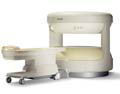
From Philips Medical Systems;
the Panorama 0.23 T, providing a new design optimized for patient comfort, faster reconstruction time than before (300 images/second) and new gradient
specifications. Philips' Panorama 0.23 T I/T supports MR-guided interventions, resulting in minimally invasive procedures, more targeted surgery, reduced recovery time and shorter hospital stays. Optional OptoGuide functionality enables real-time needle tracking. Philips' Panorama 0.23 TPanorama 0.2 R/T is the first and only open MRI system to enable radiation therapy planning using MR data sets. The Panorama also features the new and consistent Philips User Interface, an essential element of the Vequion clinical IT family of products and services.
Device Information and Specification CLINICAL APPLICATION Whole body SE, FE, IR, FFE, DEFFE, DESE, TSE, DETSE, Single shot SE, DRIVE, Balanced FFE, MRCP, Fluid Attenuated Inversion Recovery, Tur bo FLAIR, IR-TSE, T1-STIR TSE, T2-STIR TSE, Diffusion Imaging, 3D SE, 3D FFE, MTC;; Angiography: CE-ANGIO, MRA 2D, 3D TOFOpen x 46 cm x infinite (side-first patient entry) POWER REQUIREMENTS 400/480 V COOLING SYSTEM TYPE Closed loop chilled water ( chiller included) | | | |
• View the DATABASE results for 'Panorama 0.23T™' (2).
| | | | | | Further Reading: | News & More:
|
|
| |
| | | Searchterm 'Bo' was also found in the following services: | | | | |
| | |
| |
|
Device Information and Specification CLINICAL APPLICATION Whole body SE, FE, IR, STIR, FFE, DEFFE, DESE, TSE, DETSE, Single shot SE, DRIVE, Balanced FFE, MRCP, Fluid Attenuated Inversion Recovery, Tur bo FLAIR, IR-TSE, T1-STIR TSE, T2-STIR TSE, Diffusion Imaging, 3D SE, 3D FFE, Contrast Perfusion Analysis, MTC;; Angiography: CE-ANGIO, MRA 2D, 3D TOFOpen x 47 cm x infinite (side-first patient entry) POWER REQUIREMENTS 400/480 V | | | |
• View the DATABASE results for 'Panorama 0.6T™' (2).
| | | | |
| | | | | |
| |
|
From Philips Medical Systems;
this active shielded member of the Panorama product line combines the advantages of one 1.0 T system's with the possibilities of an open MRI system. The open design helps ease anxiety for claustrophobic patients and increased patient comfort whereby the field strength provides spectacular image quality and fast patient throughput.
Device Information and Specification CLINICAL APPLICATION Whole body Vertically opposed solenoids, head, head-neck, extremity, neck, body/ spine M-XL, shoulder, bilateral breast, wrist, TMJ, flex XS-S-M-L-XL-XXL SE, FE, IR, STIR, FFE, DEFFE, DESE, TSE, DETSE, Single shot SE, DRIVE, Balanced FFE, MRCP, FLAIR, Tur bo FLAIR, IR-TSE, T1-STIR TSE, T2-STIR TSE, Diffusion Imaging, 3D SE, 3D FFE, Contrast Perfusion Analysis, MTC;; Angiography: CE-ANGIO, MRA 2D, 3D TOFOpen x 47 cm x infinite (side-first patient entry) POWER REQUIREMENTS 400/480 V | | | |
• View the DATABASE results for 'Panorama 1.0T™' (2).
| | | | |
| | | | |
| | |
| | | |
|
| |
 | Look
Ups |
| |


