(SE) The most common
pulse sequence used in
MR imaging is based of the detection of a
spin or
Hahn echo. It uses 90°
radio frequency pulses to excite the
magnetization and one or more 180° pulses to refocus the spins to generate signal echoes named
spin echoes (SE).
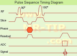
In the
pulse sequence timing diagram, the simplest form of a
spin echo sequence is illustrated.
The 90°
excitation pulse rotates the
longitudinal magnetization (
Mz) into the xy-plane and the
dephasing of the
transverse magnetization (Mxy) starts.
The following application of a 180°
refocusing pulse (rotates the
magnetization in the x-plane) generates signal echoes. The purpose of the 180° pulse is to rephase the spins, causing them to regain
coherence and thereby to recover
transverse magnetization, producing a
spin echo.
The recovery of the z-magnetization occurs with the T1
relaxation time and typically at a much slower rate than the T2-decay, because in general T1 is greater than T2 for living tissues and is in the range of 100-2000 ms.
The SE
pulse sequence was devised in the early days of
NMR days by
Carr and
Purcell and exists now in many forms: the
multi echo pulse sequence using single or multislice acquisition, the
fast spin echo (FSE/TSE)
pulse sequence,
echo planar imaging (EPI)
pulse sequence and the
gradient and spin echo (GRASE)
pulse sequence;; all are basically
spin echo sequences.
In the simplest form of SE imaging, the
pulse sequence has to be repeated as many times as the image has lines.
Contrast values:
PD weighted: Short TE (20 ms) and long TR.
T1 weighted: Short TE (10-20 ms) and short TR (300-600 ms)
T2 weighted: Long TE (greater than 60 ms) and long TR (greater than 1600 ms)
With
spin echo imaging no
T2* occurs, caused by the 180°
refocusing pulse. For this reason,
spin echo sequences are more robust against e.g.,
susceptibility artifacts than
gradient echo sequences.
See also
Pulse Sequence Timing Diagram to find a description of the components.