 | Info
Sheets |
| | | | | | | | | | | | | | | | | | | | | | | | |
 | Out-
side |
| | | | |
|
| | | 'Diffusion Weighted Sequence' | |
Result : Searchterm 'Diffusion Weighted Sequence' found in 1 term [ ] and 5 definitions [ ] and 5 definitions [ ], (+ 6 Boolean[ ], (+ 6 Boolean[ ] results ] results
| previous 6 - 10 (of 12) nextResult Pages :  [1] [1]  [2] [2]  [3] [3] |  | | |  |  |  |
| |
|
Rapid echo planar imaging and high-performance MRI gradient systems create fast-switching magnetic fields that can stimulate muscle and nerve tissues produced by either changing the electrical resistance or the potential of the excitation. There are apparently no effects on the conduction of impulses in the nerve fiber up to field strength of 0.1 T. A preliminary study has indicated neurological effects by exposition to a whole body imager at 4.0 T. Theoretical examinations argue that field strengths of 24 T are required to produce a 10% reduction of nerve impulse conduction velocity.
Nerve stimulations during MRI scans can be induced by very rapid changes of the magnetic field. This stimulation may occur for example during diffusion weighted sequences or diffusion tensor imaging and can result in muscle contractions caused by effecting motor nerves. The so-called magnetic phosphenes are attributed to magnetic field variations and may occur in a threshold field change of between 2 and 5 T/s. Phosphenes are stimulations of the optic nerve or the retina, producing a flashing light sensation in the eyes. They seem not to cause any damage in the eye or the nerve.
Varying magnetic fields are also used to stimulate bone-healing in non-unions and pseudarthroses. The reasons why pulsed magnetic fields support bone-healing are not completely understood. The mean threshold levels for various stimulations are 3600 T/s for the heart, 900 T/s for the respiratory system, and 60 T/s for the peripheral nerves.
Guidelines in the United States limit switching rates at a factor of three below the mean threshold for peripheral nerve stimulation. In the event that changes in nerve conductivity happens, the MRI scan parameters should be adjusted to reduce dB/dt for nerve stimulation. | |  | | • For this and other aspects of MRI safety see our InfoSheet about MRI Safety. | | | • Patient-related information is collected in our MRI Patient Information.
| | | | |  Further Reading: Further Reading: | | Basics:
|
|
News & More:
| |
| |
|  |  | MRI Safety Resources | | | | |
|  |  |  |
| |
|
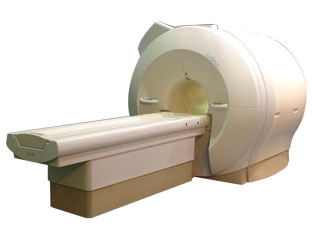
Device Information and Specification
CLINICAL APPLICATION
Whole body
CONFIGURATION
Mobile compact
Whole body, intra-operative head, neck volume, atlas head//neck vascular quadrature phased array, spine quadrature, C/T/L spine phased array, small joint, large joint, TMJ bilateral, shoulder phased array, extremity quadrature volume, wrist, hand quadrature, general purpose flexible, pelvis/abdomen phased array, body quadrature, phased array flexible, breast bilateral
IMAGING MODES
Localizer, single slice, multislice, volume
| |  | |
• View the DATABASE results for 'iMotion™ 1.5 Tesla Magnet' (2).
| | | | |
|  | |  |  |  |
| |
|

'Next generation MRI system 1.5T CHORUS developed by ISOL Technology is optimized for both clinical diagnostic imaging and for research development.
CHORUS offers the complete range of feature oriented advanced imaging techniques- for both clinical routine and research. The compact short bore magnet, the patient friendly design and the gradient technology make the innovation to new degree of perfection in magnetic resonance.'
Device Information and Specification
CLINICAL APPLICATION
Whole body
Spin Echo, Gradient Echo, Fast Spin Echo,
Inversion Recovery ( STIR, Fluid Attenuated Inversion Recovery), FLASH, FISP, PSIF, Turbo Flash ( MPRAGE ),TOF MR Angiography, Standard echo planar imaging package (SE-EPI, GE-EPI), Optional:
Advanced P.A. Imaging Package (up to 4 ch.), Advanced echo planar imaging package,
Single Shot and Diffusion Weighted EPI, IR/FLAIR EPI
STRENGTH
20 mT/m (Upto 27 mT/m)
| |  | |
• View the DATABASE results for 'CHORUS 1.5T™' (2).
| | | | |
|  | |  |  |  |
| |
|
(PWI - Perfusion Weighted Imaging) Perfusion MRI techniques (e.g. PRESTO - Principles of Echo Shifting using a Train of Observations) are sensitive to microscopic levels of blood flow. Contrast enhanced relative cerebral blood volume (rCBV) is the most used perfusion imaging.
Both, the ready availability and the T2* susceptibility effects of gadolinium, rather than the T1 shortening effects make gadolinium a suitable agent for use in perfusion imaging. Susceptibility here refers to the loss of MR signal, most marked on T2* ( gradient echo)- weighted and T2 ( spin echo)- weighted sequences, caused by the magnetic field-distorting effects of paramagnetic substances.
T2* perfusion uses dynamic sequences based on multi or single shot techniques. The T2* ( T2) MRI signal drop within or across a brain region is caused by spin dephasing during the rapid passage of contrast agent through the capillary bed. The signal decrease is used to compute the relative perfusion to that region. The bolus through the tissue is only a few seconds, high temporal resolution imaging is required to obtain sequential images during the wash in and wash out of the contrast material and therefore, resolve the first pass of the tracer. Due to the high temporal resolution, processing and calculation of hemodynamic maps are available (including mean transit time (MTT), time to peak (TTP), time of arrival (T0), negative integral (N1) and index.
An important neuroradiological indication for MRI is the evaluation of incipient or acute stroke via perfusion and diffusion imaging. Diffusion imaging can demonstrate the central effect of a stroke on the brain, whereas perfusion imaging visualizes the larger 'second ring' delineating blood flow and blood volume. Qualitative and in some instances quantitative (e.g. quantitative imaging of perfusion using a single subtraction) maps of regional organ perfusion can thus be obtained.
Echo planar and potentially echo volume techniques together with appropriate computing power offer real time images of dynamic variations in water characteristics reflecting perfusion, diffusion, oxygenation (see also Oxygen Mapping) and flow.
Another type of perfusion MR imaging allows the evaluation of myocardial ischemia during pharmacologic stress. After e.g., adenosine infusion, multiple short axis views (see cardiac axes) of the heart are obtained during the administration of gadolinium contrast. Ischemic areas show up as areas of delayed and diminished enhancement. The MRI stress perfusion has been shown to be more accurate than nuclear SPECT exams. Myocardial late enhancement and stress perfusion imaging can also be performed during the same cardiac MRI examination. | | | | | | | | | | |
• View the DATABASE results for 'Perfusion Imaging' (16).
| | |
• View the NEWS results for 'Perfusion Imaging' (3).
| | | | |  Further Reading: Further Reading: | Basics:
|
|
News & More:
| |
| |
|  | |  |  |  |
| |
|
(SENSE) A MRI technique for relevant scan time reduction. The spatial information related to the coils of a receiver array are utilized for reducing conventional Fourier encoding. In principle, SENSE can be applied to any imaging sequence and k-space trajectories. However, it is particularly feasible for Cartesian sampling schemes. In 2D Fourier imaging with common Cartesian sampling of k-space sensitivity encoding by means of a receiver array enables to reduce the number of Fourier encoding steps.
SENSE reconstruction without artifacts relies on accurate knowledge of the individual coil sensitivities. For sensitivity assessment, low-resolution, fully Fourier-encoded reference images are required, obtained with each array element and with a body coil.
The major negative point of parallel imaging techniques is that they diminish SNR in proportion to the numbers of reduction factors.
R is the factor by which the number of k-space samples is reduced. In standard Fourier imaging reducing the sampling density results in the reduction of the FOV, causing aliasing. In fact, SENSE reconstruction in the Cartesian case is efficiently performed by first creating one such aliased image for each array element using discrete Fourier transformation (DFT).
The next step then is to create a full-FOV image from the set of intermediate images. To achieve this one must undo the signal superposition underlying the fold-over effect. That is, for each pixel in the reduced FOV the signal contributions from a number of positions in the full FOV need to be separated. These positions form a Cartesian grid corresponding to the size of the reduced FOV.
The advantages are especially true for contrast-enhanced MR imaging such as
dynamic liver MRI (liver imaging) ,
3 dimensional magnetic resonance angiography (3D MRA), and magnetic resonance cholangiopancreaticography ( MRCP).
The excellent scan speed of SENSE allows for acquisition of two separate sets of hepatic MR images within the time regarded as the hepatic arterial-phase (double arterial-phase technique) as well as that of multidetector CT.
SENSE can also increase the time efficiency of spatial signal encoding in 3D MRA. With SENSE, even ultrafast (sub second) 4D MRA can be realized.
For MRCP acquisition, high-resolution 3D MRCP images can be constantly provided by SENSE. This is because SENSE resolves the presence of the severe motion artifacts due to longer acquisition time. Longer acquisition time, which results in diminishing image quality, is the greatest problem for 3D MRCP imaging.
In addition, SENSE reduces the train of gradient echoes in combination with a faster k-space traversal per unit time, thereby dramatically improving the image quality of single shot echo planar imaging (i.e. T2 weighted, diffusion weighted imaging). | |  | |
• View the DATABASE results for 'Sensitivity Encoding' (12).
| | | | |  Further Reading: Further Reading: | News & More:
|
|
| |
|  | |  |  |
|  | | |
|
| |
 | Look
Ups |
| |