| Info
Sheets |
| | | | | | | | | | | | | | | | | | | | | | | | |
 | Out-
side |
| | | | |
|
| | | | |
Result : Searchterm 'Digital' found in 3 terms [ ] and 25 definitions [ ] and 25 definitions [ ] ]
| | previous 16 - 20 (of 28) nextResult Pages : [1] [2 3 4 5 6] |  | |  | Searchterm 'Digital' was also found in the following services: | | | | |
| |  |
| |
|
'MRI system is not an expensive equipment anymore.
ENCORE developed by ISOL Technology is a low cost MRI system with the advantages like of the 1.0T MRI scanner. Developed specially for the overseas market, the ENCORE is gaining popularity in the domestic market by medium sized hospitals.
Due to the optimum RF and Gradient application technology. ENCORE enables to obtain high resolution imaging and 2D/3D Angio images which was only possible in high field MR systems.'
- Less consumption of the helium gas due to the ultra-lightweight magnet specially designed and manufactured for ISOL.
- Cost efficiency MR system due to air cooling type (equivalent to permanent magnetic).
- Patient processing speed of less than 20 minutes.'
Device Information and Specification
CLINICAL APPLICATION
Whole body
CONFIGURATION
Short bore compact
| | | | | | | | | | |
| | | | | |
| |
|
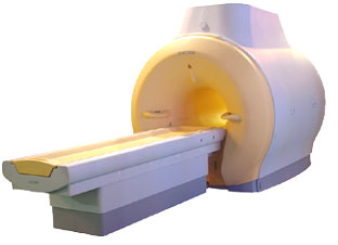
From Toshiba America Medical Systems Inc.;
FLEXART™ series is a 0.5 T superconducting MRI system that has been designed to meet the expanding role of MRI in today's clinical environment. The system utilizes innovative technologies such as digital RF, high speed actively shielded gradients and optimized RF coils which support a wide range of MRI developments.
Device Information and Specification
CLINICAL APPLICATION
Whole body
Quadrature, solenoid and multi-channel configurations
SE, FE, IR, FastSE, FastIR, FastFLAIR, Fast STIR, FastFE, FASE, Hybrid EPI, Multi Shot EPI; Angiography: 2D(gate/non-gate)/3D TOF, SORS-STC
IMAGING MODES
Single, multislice, volume study
POWER REQUIREMENTS
380/400/415/440/480 V
COOLING SYSTEM TYPE
Closed-loop water-cooled
| | | |
• View the DATABASE results for 'FLEXART™' (2).
| | | | |
| | | | | |
| |
|
Filtering deletes components of the signal, high or low frequencies, band-pass, analog or digital. Whatever pattern or algorithms can be defined for data decimation.
Low pass filtering attenuates high frequency data and passes low frequency data. The reconstructed image will look a little blurrier, but nearly similar to the original image. The blurring is caused by the fact that the high spatial frequencies are lost, which contain information about edges in the image.
High pass filtering attenuates low frequencies and passes high frequencies.
Most of the objects and contrast of the original image are lost in the reconstructed image, but the edges are clearly visible because high frequency data has been preserved. | | | |
• View the DATABASE results for 'Filtering' (8).
| | | | |  Further Reading: Further Reading: | | Basics:
|
|
News & More:
| |
| |
| | | Searchterm 'Digital' was also found in the following services: | | | | |
| | |
| |
|
| | | | | | | | |
• View the DATABASE results for 'MRI Equipment' (13).
| | |
• View the NEWS results for 'MRI Equipment' (4).
| | | | | | Further Reading: | News & More:
|
|
| |
| | | | | |
| |
|
Device Information and Specification CLINICAL APPLICATION Whole body Quadrature, solenoid and multi-channel configurations SE, FE, IR, FastSE, FastIR, FastFLAIR, Fast STIR, FastFE, FASE, Hybrid EPI, Multi Shot EPI; Angiography: 2D(gate/non-gate)/3D TOF, SORS-STC IMAGING MODES Single, multislice, volume study POWER REQUIREMENTS 380/400/415/440/480 V COOLING SYSTEM TYPE Cryogenless | | | |
• View the DATABASE results for 'OPART™' (2).
| | | | |
| | | | |
| | |
| | | |
|
| |
 | Look
Ups |
| |