Magnetic Resonance - Technology Information Portal Welcome to MRI Technology
Info
  Sheets

Out-
      side


 'Dual Spin Echo' 
SEARCH FOR    
 
  2 3 5 A B C D E F G H I J K L M N O P Q R S T U V W X Y Z
Result : Searchterm 'Dual Spin Echo' found in 0 term [] and 0 definition [], (+ 5 Boolean[] results
1 - 5 (of 5)     
Result Pages : [1]
Searchterm 'Dual Spin Echo' was also found in the following service: 
spacer
Forum  (1)  
 
Dual Echo Fast Acquisition Interleaved Spin EchoInfoSheet: - Sequences - 
Intro, 
Overview, 
Types of, 
etc.
 
(DEFAISE) Simultaneously acquired T2 and density weighted echoes in a FSE sequence.

See Fast Spin Echo.
spacer
 
• Share the entry 'Dual Echo Fast Acquisition Interleaved Spin Echo':  Facebook  Twitter  LinkedIn  
 
Further Reading:
  Basics:
BASIC PRINCIPLES OF MR IMAGING
   by spinwarp.ucsd.edu    
MRI Resources 
Contrast Enhanced MRI - Fluorescence - Pediatric and Fetal MRI - Manufacturers - Devices - RIS
 
Fast Spin EchoForum -
related threadsInfoSheet: - Sequences - 
Intro, 
Overview, 
Types of, 
etc.
 
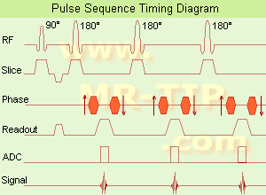
Fast Spin Echo Diagram (FSE) In the pulse sequence timing diagram, a fast spin echo sequence with an echo train length of 3 is illustrated. This sequence is characterized by a series of rapidly applied 180° rephasing pulses and multiple echoes, changing the phase encoding gradient for each echo.
The echo time TE may vary from echo to echo in the echo train. The echoes in the center of the K-space (in the case of linear k-space acquisition) mainly produce the type of image contrast, whereas the periphery of K-space determines the spatial resolution. For example, in the middle of K-space the late echoes of T2 weighted images are encoded. T1 or PD contrast is produced from the early echoes.
The benefit of this technique is that the scan duration with, e.g. a turbo spin echo turbo factor / echo train length of 9, is one ninth of the time. In T1 weighted and proton density weighted sequences, there is a limit to how large the ETL can be (e.g. a usual ETL for T1 weighted images is between 3 and 7). The use of large echo train lengths with short TE results in blurring and loss of contrast. For this reason, T2 weighted imaging profits most from this technique.
In T2 weighted FSE images, both water and fat are hyperintense. This is because the succession of 180° RF pulses reduces the spin spin interactions in fat and increases its T2 decay time. Fast spin echo (FSE) sequences have replaced conventional T2 weighted spin echo sequences for most clinical applications. Fast spin echo allows reduced acquisition times and enables T2 weighted breath hold imaging, e.g. for applications in the upper abdomen.
In case of the acquisition of 2 echoes this type of a sequence is named double fast spin echo / dual echo sequence, the first echo is usually density and the second echo is T2 weighted image. Fast spin echo images are more T2 weighted, which makes it difficult to obtain true proton density weighted images. For dual echo imaging with density weighting, the TR should be kept between 2000 - 2400 msec with a short ETL (e.g., 4).
Other terms for this technique are:
Turbo Spin Echo
Rapid Imaging Spin Echo,
Rapid Spin Echo,
Rapid Acquisition Spin Echo,
Rapid Acquisition with Refocused Echoes
 
Images, Movies, Sliders:
 Lumbar Spine T2 FSE Sagittal  Open this link in a new window
    

Courtesy of  Robert R. Edelman
 MRI - Anatomic Imaging of the Foot  Open this link in a new window
    
SlidersSliders Overview

 Lumbar Spine T2 FSE Axial  Open this link in a new window
    

Courtesy of  Robert R. Edelman
 
spacer

• View the DATABASE results for 'Fast Spin Echo' (31).Open this link in a new window

 
Further Reading:
  Basics:
MYELIN-SELECTIVE MRI: PULSE SEQUENCE DESIGN AND OPTIMIZATION
   by www.imaging.robarts.ca    
Advances in Magnetic Resonance Neuroimaging
Friday, 27 February 2009   by www.ncbi.nlm.nih.gov    
  News & More:
New MR sequence helps radiologists more accurately evaluate abnormalities of the uterus and ovaries
Thursday, 23 April 2009   by www.eurekalert.org    
Spin echoes, CPMG and T2 relaxation - Introductory NMR & MRI from Magritek
2013   by www.azom.com    
MRI Resources 
Process Analysis - MRCP - Research Labs - NMR - Cochlear Implant - MRI Centers
 
Dual Echo Fast Gradient EchoInfoSheet: - Sequences - 
Intro, 
Overview, 
Types of, 
etc.
 
(DE FGRE, Dual/FFE, DE FFE) Simultaneously acquired in and out of phase TE gradient echo images. To quantitatively measure the signal intensity differences between out of phase and in phase images the parameters should be the same except for the TE.
The chemical shift artifact appearing on the out-of-phase image allows for the detection of lipids in the liver or adrenal gland, such as diffuse fatty infiltration, focal fatty infiltration, focal fatty sparing, benign adrenocortical masses and intracellular lipids within a hepatocellar neoplasm, where spin echo and fat suppression techniques are not as sensitive. Specific pathologies that have been reported include liver lipoma, angiomyolipoma, myelolipoma, metastatic liposarcoma, teratocarcinoma, melanoma, haemorrhagic neoplasm and metastatic choriocarcinoma.
 
Images, Movies, Sliders:
 MRI Liver In Phase  Open this link in a new window
    
 MRI Liver Out Of Phase  Open this link in a new window
    
 
spacer

• View the DATABASE results for 'Dual Echo Fast Gradient Echo' (2).Open this link in a new window

 
Further Reading:
  News & More:
Adrenal Myelolipoma
Tuesday, 19 June 2001   by www.emedicine.com    
Searchterm 'Dual Spin Echo' was also found in the following service: 
spacer
Forum  (1)  
 
Free Induction Decay
 
(FID) A free induction decay curve is generated as excited nuclei relax. The amplitude of the FID signal becomes smaller over time as net magnetization returns to equilibrium. If transverse magnetization of the spins is produced, e.g. by a 90° pulse, a transient MR signal will result that will decay toward zero with a characteristic time constant T2 (or T2*); this decaying signal is the free induction decay.
The signal peaks of the echoes fall onto this T2 decay curve, while at each echo the signals arise and decay with T2*. The typical T2 relaxation times being of the order of 5-200 ms in the human body. The first part of the FID is not observable (named the 'receiver dead time') caused by residual effects of the powerful exciting radio frequency pulse on the electronics of the receiver.
spacer

• View the DATABASE results for 'Free Induction Decay' (8).Open this link in a new window

 
Further Reading:
  Basics:
Free induction decay
   by en.wikipedia.org    
  News & More:
Magnetic resonance imaging
   by www.scholarpedia.org    
MRI Resources 
Musculoskeletal and Joint MRI - Mobile MRI - Databases - Shielding - Knee MRI - Distributors
 
Incoherent Gradient Echo (RF Spoiled)InfoSheet: - Sequences - 
Intro, 
Overview, 
Types of, 
etc.
 
A gradient echo is generated by using a pair of bipolar gradient pulses. The gradient field is negatively pulsed, causing the spins of the xy-magnetization to dephase. A second gradient pulse is applied with the opposite polarity. During the pulsing, the spins that dephased begin to rephase and generate a gradient echo.
Spoiling can be accomplished by RF or a gradient. The incoherent RF spoiled type of a gradient echo sequence use a continuous shifting of the RF pulse to spoil the residual transverse magnetization. The phase of the RF excitation and receiver channel are varied pseudo randomly with each excitation cycle to prevent the xy magnetization from achieving steady state. T2* does not dominate image contrast, so T1 and PD weighting is practical. This method is effective and can be used to achieve a shorter TR, due to a lack of additional gradients. Spoiling eliminates the effect of the remaining xy-magnetization and leads to steady state longitudinal magnetization. These sequences can be used for breath hold, dynamic imaging and in cine and volume acquisitions.
spacer
MRI Resources 
Image Quality - Functional MRI - Blood Flow Imaging - Crystallography - Knee MRI - Collections
 
     1 - 5 (of 5)     
Result Pages : [1]
 Random Page
 
Share This Page
FacebookTwitterLinkedIn

MR-TIP    
Community   
User
Pass
Forgot your UserID/Password ?    


MRI is trending to low field magnets :
reduced costs will lead to this change 
AI will close the gap to high field 
only in remote areas 
is only temporary 
never 

Look
      Ups


MR-TIP.com uses cookies! By browsing MR-TIP.com, you agree to our use of cookies.

Magnetic Resonance - Technology Information Portal
Member of SoftWays' Medical Imaging Group - MR-TIP • Radiology-TIP • Medical-Ultrasound-Imaging • 
Copyright © 2003 - 2024 SoftWays. All rights reserved. [ 4 December 2024]
Terms of Use | Privacy Policy | Advertising
 [last update: 2024-02-26 03:41:00]