Magnetic Resonance - Technology Information Portal Welcome to MRI Technology
Info
  Sheets

Out-
      side


 'Fat Suppression' 
SEARCH FOR    
 
  2 3 5 A B C D E F G H I J K L M N O P Q R S T U V W X Y Z
Result : Searchterm 'Fat Suppression' found in 2 terms [] and 27 definitions []
previous     11 - 15 (of 29)     next
Result Pages : [1]  [2 3 4 5 6]
Searchterm 'Fat Suppression' was also found in the following services: 
spacer
News  (1)  Resources  (2)  Forum  (6)  
 
Aurora® 1.5T Dedicated Breast MRI SystemInfoSheet: - Devices -
Intro, 
Types of Magnets, 
Overview, 
etc.
 
www.auroramri.com/mri/product/ From Aurora Imaging Technology, Inc.;
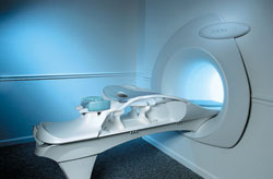
The Aurora® 1.5T Dedicated Breast MRI System with Bilateral SpiralRODEO™ is the first and only FDA approved MRI device designed specifically for breast imaging. The Aurora System, which is already in clinical use at a growing number of leading breast care centers in the US, Europe, got in December 2006 also the approval from the State Food and Drug Administration of the People's Republic of China (SFDA).
'Some of the proprietary and distinguishing features of the Aurora System include: 1) an ellipsoid magnetic shim that provides coverage of both breasts, the chest wall and bilateral axillary lymph nodes; 2) a precision gradient coil with the high linearity required for high resolution spiral reconstruction;; 3) a patient-handling table that provides patient comfort and procedural utility; 4) a fully integrated Interventional System for MRI guided biopsy and localization; and 5) the user-friendly AuroraCADâ„¢ computer-aided image display system designed to improve the accuracy and efficiency of diagnostic interpretations.'
Device Information and Specification
CLINICAL APPLICATION
CONFIGURATION
Short bore compact
IMAGING MODES
Bi-lateral RODEO fat suppression, high resolution Rotating Delivery of Excitation Offresonance Spiral, integrated targeting SW compatible with major MR guided intervention equipment
TR
10 ms for gradient echo and less than 2,500 ms for T2 weighted spin echo
TE
From 5 ms for RODEO Plus to over 80 ms, 120 ms for T2 sequences
SINGLE/MULTI SLICE
Around 0.02 sec for a 256x256 image, 12.4 sec for a 512 x 512 x 32 multislice set
FOV
20 - 36 cm, max. elliptical 36 x 44 cm
MEASURING MATRIX
512 x 512
BORE DIAMETER
or W x H
64 cm diameter (gantry)
MAGNET WEIGHT
8,500 lbs
H*W*D
240 x 188 x 163 cm
POWER REQUIREMENTS
150A/120V-208Y/3 Phase//60 Hz/5 Wire
COOLING SYSTEM
Helium for magnet, distilled/de-ionized water for coil;
spacer
 
• Related Searches:
    • Breast MRI
    • MRI Equipment
    • MRI
    • Hardware
    • Superconducting Magnet
 
Further Reading:
  News & More:
Aurora Imaging Technology Announces Approval in China
   by salesandmarketingnetwork.com    
Aurora Imaging Technology, Inc. Release: Results of a Multi-Center Trial Demonstrates Superior Diagnostic Performance of the Aurora® 1.5T Dedicated Breast MRI System Over Whole-Body Breast MRI
Monday, 1 October 2012   by www.biospace.com    
MRI Resources 
Mass Spectrometry - DICOM - Jobs pool - Chemistry - Resources -
 
Bilateral Breast Imaging in the Sagittal View with SENSEInfoSheet: - Sequences - 
Intro, 
Overview, 
Types of, 
etc.
 
(BLISS) BLISS is a magnetic resonance imaging technique that provides the measurement of two bilateral volumes in a single acquisition. Localized shimming for each volume improves the fat suppression. BLISS is an imaging sequence for sagittal breast MRI studies with high spatial resolution and short scan time.
spacer
MRI Resources 
MR Myelography - Homepages - Examinations - Musculoskeletal and Joint MRI - Education pool - Implant and Prosthesis
 
Black Boundary ArtifactInfoSheet: - Artifacts - 
Case Studies, 
Reduction Index, 
etc.MRI Resource Directory:
 - Artifacts -
 
Quick Overview
Please note that there are different common names for this artifact.
Artifact Information
NAME
Black boundary, dark boundary, contour, chemical shift, relief
DESCRIPTION
Black contours at boundaries
Black boundary artifacts are black lines following voxels where both water and fat protons are present in the same voxel. This artifact arise along the boundary of organs or tissues perpendicular to the frequency encoding direction, and occurs preferentially in gradient echo sequences with out of phase echo times.
mri safety guidance
Image Guidance
Fat suppression techniques eliminate this artifact. For artifact reducing helps a smaller water fat shift (high bandwidth), a higher matrix or/and an in phase TE.

See also Chemical Shift Artifact.
spacer

• View the DATABASE results for 'Black Boundary Artifact' (4).Open this link in a new window

 
Further Reading:
  Basics:
What is chemical shift artefact? Why does it occur? How many Hz at 1.5 T?
   by www.revisemri.com    
Searchterm 'Fat Suppression' was also found in the following services: 
spacer
News  (1)  Resources  (2)  Forum  (6)  
 
Chemical Shift Selective Imaging SequenceInfoSheet: - Sequences - 
Intro, 
Overview, 
Types of, 
etc.MRI Resource Directory:
 - Spectroscopy pool -
 
(CHESS) A sequence for water suppression in proton MR spectroscopy and for water or fat suppression in MR imaging. This technique uses a frequency-selective 90° pulse to selectively excite the water signal, followed by a spoiler gradient to dephase the resulting magnetization. The gradients may be repeated several times in different directions to increase its effectiveness.

See also Chemical Shift Imaging and Chemical Shift.
spacer

• View the DATABASE results for 'Chemical Shift Selective Imaging Sequence' (2).Open this link in a new window

MRI Resources 
Quality Advice - Contrast Enhanced MRI - Spine MRI - Stimulator pool - Education - MRI Training Courses
 
Dual Echo Fast Gradient EchoInfoSheet: - Sequences - 
Intro, 
Overview, 
Types of, 
etc.
 
(DE FGRE, Dual/FFE, DE FFE) Simultaneously acquired in and out of phase TE gradient echo images. To quantitatively measure the signal intensity differences between out of phase and in phase images the parameters should be the same except for the TE.
The chemical shift artifact appearing on the out-of-phase image allows for the detection of lipids in the liver or adrenal gland, such as diffuse fatty infiltration, focal fatty infiltration, focal fatty sparing, benign adrenocortical masses and intracellular lipids within a hepatocellar neoplasm, where spin echo and fat suppression techniques are not as sensitive. Specific pathologies that have been reported include liver lipoma, angiomyolipoma, myelolipoma, metastatic liposarcoma, teratocarcinoma, melanoma, haemorrhagic neoplasm and metastatic choriocarcinoma.
 
Images, Movies, Sliders:
 MRI Liver In Phase  Open this link in a new window
    
 MRI Liver Out Of Phase  Open this link in a new window
    
 
spacer

• View the DATABASE results for 'Dual Echo Fast Gradient Echo' (2).Open this link in a new window

 
Further Reading:
  News & More:
Adrenal Myelolipoma
Tuesday, 19 June 2001   by www.emedicine.com    
MRI Resources 
MRI Training Courses - Case Studies - Nerve Stimulator - Portals - Non-English - Open Directory Project
 
previous      11 - 15 (of 29)     next
Result Pages : [1]  [2 3 4 5 6]
 Random Page
 
Share This Page
FacebookTwitterLinkedIn

MR-TIP    
Community   
User
Pass
Forgot your UserID/Password ?    


New acceleration techniques will :
reduce scan times 
cause artifacts 
increase expenses 
be useful if you have a lot of experience 
doesn't do much 
never heard of 

Look
      Ups


MR-TIP.com uses cookies! By browsing MR-TIP.com, you agree to our use of cookies.

Magnetic Resonance - Technology Information Portal
Member of SoftWays' Medical Imaging Group - MR-TIP • Radiology-TIP • Medical-Ultrasound-Imaging • 
Copyright © 2003 - 2024 SoftWays. All rights reserved. [ 21 November 2024]
Terms of Use | Privacy Policy | Advertising
 [last update: 2024-02-26 03:41:00]