Magnetic Resonance - Technology Information Portal Welcome to MRI Technology
Info
  Sheets

Out-
      side


 'HIS' 
SEARCH FOR    
 
  2 3 5 A B C D E F G H I J K L M N O P Q R S T U V W X Y Z
Result : Searchterm 'HIS' found in 1 term [] and 429 definitions []
previous     46 - 50 (of 430)     next
Result Pages : [1]  [2 3 4 5 6 7 8 9 10 11 12 13 14 15 16 17 18 19 20 ... ]
Searchterm 'HIS' was also found in the following services: 
spacer
News  (375)  Resources  (57)  Forum  (303)  
 
Berlex Laboratories, Inc.MRI Resource Directory:
 - Manufacturers -
 
www.berleximaging.com [This entry is marked for removal.]

Berlex Laboratories, Inc. was an US affiliate of Schering AG Germany (now Bayer Schering Pharma AG ).
In June 2006 Bayer AG has bought the majority of shares of Schering AG. As a result of this take-over the new formed company has been named Bayer HealthCare Pharmaceuticals.
For contrast agents distributed by Berlex Laboratories, Inc. please see Bayer HealthCare Pharmaceuticals.
spacer

• View the NEWS results for 'Berlex Laboratories, Inc.' (1).Open this link in a new window.
MRI Resources 
Shoulder MRI - Safety Products - Image Quality - Collections - Societies - Veterinary MRI
 
Bipolar Gradient Pulse
 
Bipolar gradients are two gradients with the same magnitude but opposite gradient direction. A bipolar gradient pulse is produced if one of the bipolar gradients is switched e.g., in negative direction and then switched in the opposite direction for an equivalent amount of time.
Bipolar gradients are used e.g. in phase contrast and diffusion weighted sequences. A bipolar gradient pulse pair produces a phase shift, which depends on the velocity component along this gradient. Motion along a bipolar gradient pulse pair results in a flow-induced phase shift of the transverse magnetization. The bipolar gradient pulse pair will not affect stationary spins. The amount of phase shifts depends on the area of each gradient pulse, and distance between the pulses. An echo occurring after such a gradient is flow compensated for velocity. A slight shift in the balance of this gradient will introduce a defined flow sensitivity of the pulse sequence.
 
Images, Movies, Sliders:
 PCA-MRA 3D Brain Venography Colored MIP  Open this link in a new window


spacer

• View the DATABASE results for 'Bipolar Gradient Pulse' (7).Open this link in a new window

MRI Resources 
Claustrophobia - Libraries - Spectroscopy pool - Lung Imaging - MRI Reimbursement - Health
 
Bird Cage CoilInfoSheet: - Coils - 
Intro, 
Overview, 
etc.MRI Resource Directory:
 - Coils -
 
A RF coil, often a transmit receive coil with a number of wires running along the z-direction, arranged to give a cosine current variation around the circumference of the coil, which looks like a bird cage. The bird cage coil works on a different principle to conventionally tuned local and surround coils in that it behaves like a tuned transmission line with one complete cycle of standing wave around the circumference. The frequency supply is generated by an oscillator, which is modulated to form a shaped pulse by a product detector controlled by the waveform generator. The signal must be amplified to 1000's of watts. This can be done using either solid state electronics, valves or a combination of both.
The bird cage coil design provides the best field homogeneity of all RF imaging coils.
One advantage is that it is simple to produce an exceedingly uniform B1 radio frequency field over most of the coil's volume, with the result of images with a high degree of uniformity.
A second advantage is that nodes with zero voltage occur 90° away from the driven part of the coil, thus facilitating the introduction of a second signal in quadrature, which produces a circularly polarized radio frequency field.
This type of volume coil is used for brain (head) MRI, or MR imaging of joints, such as the wrist or knees.

See also the related poll result: '3rd party coils are better than the original manufacturer coils'
 
Images, Movies, Sliders:
 Brain MRI Images T1  Open this link in a new window
 Anatomic MRI of the Knee 1  Open this link in a new window
    
SlidersSliders Overview

 MRI of the Brain Stem with Temoral Bone and Auditory System  Open this link in a new window
    
SlidersSliders Overview

 
spacer

• View the DATABASE results for 'Bird Cage Coil' (4).Open this link in a new window

 
Further Reading:
  Basics:
HEAD AND NECK MRI GRADIENT COIL DESIGN
Thursday, 26 August 1999   by www.imaging.robarts.ca    
System Architecture
2003   by www.revisemri.com    
  News & More:
On the Horizon - Next Generation MRI
Wednesday, 23 October 2013   by thefutureofthings.com    
TOSHIBA INTRODUCES 32 ELEMENT COILS FOR ITS VANTAGE TITAN MR SYSTEMS
Tuesday, 9 November 2010   by medical.toshiba.com    
Searchterm 'HIS' was also found in the following services: 
spacer
News  (375)  Resources  (57)  Forum  (303)  
 
Blood Pool AgentsInfoSheet: - Contrast Agents - 
Intro, Overview, 
Characteristics, 
Types of, 
etc.MRI Resource Directory:
 - Contrast Agents -
 
Blood pool agents (intravascular contrast agents) remain in the blood for a prolonged time compared with conventional contrast agents, which diffuse quickly into the interstitial space. Magnetic resonance angiography (MRA), cardiovascular imaging, or contrast enhanced MRIs are possible over an hour or more. This advantage over conventional MRI contrast media allows also higher resolution MRA of several territories using respiratory or cardiac gating techniques with a single contrast bolus.
Different types of blood pool contrast agents:
Blood pool MRI contrast agents with their longer intravascular circulation can be designed to be targeted to necrotic myocardium, to assess myocardial viability, or tumor directed to provide better diagnostic information for various tumors. A disadvantage of the use of blood pool agents for MRA is that the separation of arteries and veins is more difficult because they are present in both and the overlapping of those vessels is disturbing. This can be solved by e.g. different MIP segmentation algorithms.

See also Necrosis Avid Contrast Agent, Tumor Specific Agents, Feruglose, Gadofosveset Trisodium (Vasovist), Ultrasmall Superparamagnetic Iron Oxide and Contrast Medium.
spacer

• View the DATABASE results for 'Blood Pool Agents' (16).Open this link in a new window


• View the NEWS results for 'Blood Pool Agents' (1).Open this link in a new window.
 
Further Reading:
  Basics:
Ablavar Prescribing Information
   by www.ablavar.com    
Lantheus Medical Imaging, Inc. Launches ABLAVAR™ (Gadofosveset Trisodium), a New Diagnostic Magnetic Resonance Angiography Agent
Wednesday, 20 January 2010   by www.radiopharm.com    
Blood-Pool Imaging Using Technetium-99m-Labeled Liposomes(.pdf)
   by jnm.snmjournals.org    
  News & More:
Multimodal Nanoparticles for Quantitative Imaging(.pdf)
Tuesday, 13 December 2011   by alexandria.tue.nl    
MAGNETIC RESONANCE IMAGING OF FOCAL LIVER LESIONS(.pdf)
2002
MRI Resources 
NMR - Jobs - Distributors - Abdominal Imaging - MRI Training Courses - Cochlear Implant
 
C-SCAN™InfoSheet: - Devices -
Intro, 
Types of Magnets, 
Overview, 
etc.MRI Resource Directory:
 - Devices -
 
www.gehealthcare.com/usen/mr/cscan/index.html Manufactured by Esaote S.p.A.; compact in-office MRI system, fits in a 90 ft2 (8.4 m2) space and requires no shielding or special power. This low field MRI magnet is optimized for orthopedic use and imaging of the extremities.
The C-SCAN™ is developed from the ARTOSCAN™ - M with a new computer platform, and is also known as Artoscan C.
Esaote North America and Hologic Inc. are the U.S. distributors of this MRI device.
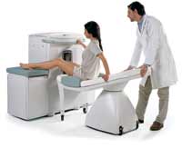
Device Information and Specification
CLINICAL APPLICATION
Dedicated extremity
CONFIGURATION
Closed
Linear and dual phased array, knee, ankle, wrist (6 total) coils
PULSE SEQUENCES
SE, GE, IR, STIR, FSE, 3D CE, GE-STIR, 3D GE, ME, TME, HSE
IMAGING MODES
Single, multislice, volume study, fast scan, multi slab
TR
12 - 5,000 msec
TE
5 - 220 msec
SINGLE SLICE
0.8 sec/image
MULTISLICE
0.8 sec/image
FOV
10 - 20 cm
2D: 2 mm - 10 mm;
3D: 0.6 mm - 10 mm
512 x 512
MEASURING MATRIX
256 x 256 maximum
PIXEL INTENSITY
4,096 gray lvls, 256 lvls in 3D
MAGNET TYPE
Permanent
BORE DIAMETER
or W x H
33.6 x 16 cm
MAGNET WEIGHT
960 kg
H*W*D
124 x 76 x 60 cm
POWER REQUIREMENTS
100/110/200/220/230/240
STRENGTH
10 mT/m
5-GAUSS FRINGE FIELD, radial/axial
28 cm/60 cm
Passive
spacer

• View the DATABASE results for 'C-SCAN™' (4).Open this link in a new window

MRI Resources 
Shielding - Examinations - Universities - Devices - MR Myelography - PACS
 
previous      46 - 50 (of 430)     next
Result Pages : [1]  [2 3 4 5 6 7 8 9 10 11 12 13 14 15 16 17 18 19 20 ... ]
 Random Page
 
Share This Page
FacebookTwitterLinkedIn

MR-TIP    
Community   
User
Pass
Forgot your UserID/Password ?    


New acceleration techniques will :
reduce scan times 
cause artifacts 
increase expenses 
be useful if you have a lot of experience 
doesn't do much 
never heard of 

Look
      Ups


MR-TIP.com uses cookies! By browsing MR-TIP.com, you agree to our use of cookies.

Magnetic Resonance - Technology Information Portal
Member of SoftWays' Medical Imaging Group - MR-TIP • Radiology-TIP • Medical-Ultrasound-Imaging • 
Copyright © 2003 - 2024 SoftWays. All rights reserved. [ 18 September 2024]
Terms of Use | Privacy Policy | Advertising
 [last update: 2024-02-26 03:41:00]