 | Info
Sheets |
| | | | | | | | | | | | | | | | | | | | | | | | |
 | Out-
side |
| | | | |
|
| | | | | |  | Searchterm 'HIS' was also found in the following services: | | | | |
|  |  |
| |
|
(ISIS) Image selected in vivo spectroscopy is used as a localization sequence to provide complete gradient controlled three-dimensional localization with a reduced number of sequence cycles, e.g. for in vivo 31P spectroscopy.
The ISIS method generates three 180° pulses prior to a 90° pulse, after which the free induction decay is recorded. Specific 180° pulses (slice-selective) are combined and the FID's added or subtracted to generate a spectrum.
An advantage of the ISIS method is that the magnetization (before the final 90° pulse) is predominantly along the z-axis and so T2 effects are relatively small. T his explains the value of t his technique for 31P data acquisition, because some phosphorus metabolites (e.g. ATP) have short T2 values.
A disadvantage is that eight acquisitions are required to accomplish the spatial localization, therefore the sequence cannot be used for localized shimming.
Another problem, because any variation between these data collections (for example, due to movement) will degrade these applications, can be solved by incorporating outer volume suppression techniques such as OSIRIS (modified ISIS). | | | | | |
| | | | | |
| |
|
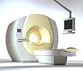
From Philips Medical Systems;
the Intera-family offers with t his member a wide range of possibilities, efficiency and a ergonomic and intuitive serving-platform. Also available as Intera CV for cardiac and Intera I/T for interventional MR procedures.
The scanners are also equipped with SENSE technology, which is essential for high-quality contrast enhanced magnetic resonance angiography, interactive cardiac MR and diffusion tensor imaging ( DTI) fiber tracking.
The increased accuracy and clarity of MR scans obtained with t his technology allow for faster and more accurate diagnosis of potential problems like patient friendliness and expands the breadth of applications including cardiology, oncology and interventional MR.
Device Information and Specification
CLINICAL APPLICATION
Whole body
CONFIGURATION
Short bore compact
Standard: head, body, C1, C3; Optional: Small joint, flex-E, flex-R, endocavitary (L and S), dual TMJ, knee, neck, T/L spine, breast; Optional phased array: Spine, pediatric, 3rd party connector; Optional SENSE coils: Flex-S-M-L, flex body, flex cardiac
SE, Modified-SE ( TSE), IR (T1, T2, PD), STIR, FLAIR, SPIR, FFE, T1-FFE, T2-FFE, Balanced FFE, TFE, Balanced TFE, Dynamic, Keyhole, 3D, Multi Chunk 3D, Multi Stack 3D, K Space Shutter, MTC, TSE, Dual IR, DRIVE, EPI, Cine, 2DMSS, DAVE, Mixed Mode; Angiography: PCA, MCA, Inflow MRA, CE
TR
2.9 (Omni), 1.6 (Power), 1.6 (Master/Expl) msec
TE
1.0 (Omni), 0.7 (Power), 0.5 (Master/Expl) msec
RapidView Recon. greater than 500 @ 256 Matrix
0.1 mm(Omni), 0.05 mm (Pwr/Mstr/Expl)
128 x 128, 256 x 256,512 x 512,1024 x 1024 (64 for BOLD img.)
Variable in 1% increments
Lum.: 120 cd/m2; contrast: 150:1
Variable (op. param. depend.)
POWER REQUIREMENTS
380/400 V
| | | |
• View the DATABASE results for 'Intera 1.5T™' (2).
| | | | |
| | | | | |
| |
|
(IVIM) Spins moving in fluids with different velocities and possibly in different directions.
T his is being found to a small degree in all tissues as a result of capillary perfusion or diffusion. Important velocity changes occur as one moves from the vessel wall towards the center of the vessel.
Hence, spins (to a variable degree) have different velocities within a single imaging voxel.
T his effect can be measured using special pulse sequences such as in diffusion imaging or diffusion weighed imaging. When the velocity differences are marked, as occurs in larger blood vessels, effects due to IVIM are visible in standard MR images and give rise to flow related dephasing. The effects are more visible when longer echo times are used. | | | |
• View the DATABASE results for 'Intravoxel Incoherent Motion' (3).
| | | | |  Further Reading: Further Reading: | | Basics:
|
|
News & More:
| |
| |
| | | Searchterm 'HIS' was also found in the following services: | | | | |
| | |
| |
|
| | | | | |
• View the DATABASE results for 'Ionic Intravenous Contrast Agents' (5).
| | | | | | Further Reading: | Basics:
|
|
News & More:
| |
| |
| | | | | |
| |
|
| | | |
• View the DATABASE results for 'MR Guided Interventions' (8).
| | | | | | Further Reading: | Basics:
|
|
News & More:
|  |
AI analysis finds younger AFib patients benefit from MRI-guided ablation treatments
Friday, 25 August 2023 by www.eurekalert.org | | |
Theranostic nano-platform for MRI-guided synergistic therapy against breast cancer
Monday, 26 September 2022 by phys.org | | |
Magnetic seeds used to heat and kill cancer
Tuesday, 1 February 2022 by www.sciencedaily.com | | |
What is the effect of MRI with targeted biopsies on the rate of patients discontinuing active surveillance? A reflection of the use of MRI in the PRIAS study
Thursday, 8 April 2021 by www.docwirenews.com | | |
Modeling of Active Shimming of Metallic Needles for Interventional MRI
Monday, 29 June 2020 by pubmed.ncbi.nlm.nih.gov | | |
Magnetic Resonance Imaging Guided Confirmatory Biopsy for Initiating Active Surveillance of Prostate Cancer
Wednesday, 11 September 2019 by jamanetwork.com | | |
FDA clears ViewRay's next-gen, MRI-guided radiation therapy device
Tuesday, 28 February 2017 by www.fiercebiotech.com | | |
Siemens, U. of Twente Biopsy Robot Promises Greater Precision, Less Cost
Friday, 22 January 2016 by www.meddeviceonline.com | | |
Magnetic resonance-guided motorized transcranial ultrasound system for blood-brain barrier permeabilization along arbitrary trajectories in rodents
Thursday, 24 December 2015 by www.ncbi.nlm.nih.gov | | |
New MRI-Guided Catheter Shows Major Potential for Stroke Treatment
Tuesday, 29 December 2015 by www.radiology.ucsf.edu | | |
Polish study on MRI-ultrasound for targeted prostate biopsy wins CEM award
Tuesday, 12 November 2013 by medicalxpress.com | | |
C4 Imaging Announces FDA 510(k) Clearance of its Positive-Signal MRI Marker - Sirius™
Friday, 6 December 2013 by www.digitaljournal.com |
|
| |
| | | | |
| | | |
|
| |
 | Look
Ups |
| |


