Magnetic Resonance - Technology Information Portal Welcome to MRI Technology
Info
  Sheets

Out-
      side


 'Half Scan' 
SEARCH FOR    
 
  2 3 5 A B C D E F G H I J K L M N O P Q R S T U V W X Y Z
Result : Searchterm 'Half Scan' found in 1 term [] and 3 definitions [], (+ 8 Boolean[] results
1 - 5 (of 12)     next
Result Pages : [1]  [2 3]
Searchterm 'Half Scan' was also found in the following service: 
spacer
News  (3)  
 
Half Scan
 
(HS) A method in which approximately one half of the acquisition matrix in the phase encoding direction is acquired. Half scan is possible because of symmetry in acquired data. Since negative values of phase encoded measurements are identical to corresponding positive values, only a little over half (more than 62.5%) of a scan actually needs to be acquired to replicate an entire scan. This results in a reduction in scan time at the expense of signal to noise ratio. The time reduction can be nearly a factor of two, but full resolution is maintained.
Half scan can be used when scan times are long, the signal to noise ratio is not critical and where full spatial resolution is required. Half scan is particularly appropriate for scans with a large field of view and relatively thick slices; and, in 3D scans with many slices. In some fast scanning techniques the use of Half scan enables a shorter TE thus improving contrast. For this reason, the Half scan parameter is located in the contrast menu.

More information about scan time reduction; see also partial fourier technique.
spacer
 
• Share the entry 'Half Scan':  Facebook  Twitter  LinkedIn  
MRI Resources 
Developers - Universities - Intraoperative MRI - Equipment - Safety Training - Stent
 
Fractional Nex Imaging
 
Fractional Nex imaging (GE Healthcare term for imaging with a Nex value less than 1) benefits from the conjugate symmetry of the k-space to reduce the number of phase encoding acquisitions. With fractional Nex imaging (similar to partial Fourier or Half Scan), just over half of the data are acquired and the data from the lower part of k-space are used to fill the upper part, without sampling the upper part. Fractional Nex imaging sequences use a number of excitations values between 0.5 and 1. These values are a bit misleading, because the number of phase encoding steps is reduced, and not the NEX.
Fractional Nex imaging reduces the scan time considerable, by preserving the same contrast between the tissues. The effect by acquiring fewer data points is that the signal to noise ratio decreases.

See also acronyms for 'partial averaging//fractional Nex imaging' from different manufacturers.
spacer
 
Further Reading:
  Basics:
Method and apparatus for subterranean formation flow imaging
   by www.google.com    
CHAPTER-12
   by www.cis.rit.edu    
  News & More:
A Practical Guide to Cardiovascular MRI
   by www.gehealthcare.com    
MRI Resources 
MRI Reimbursement - Manufacturers - Software - Education pool - Nerve Stimulator - Developers
 
O-SCAN™InfoSheet: - Devices -
Intro, 
Types of Magnets, 
Overview, 
etc.
 
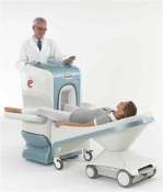
www.fonar.com/standup.htm www.fonar.com/standup.htm O-scan is manufactured and distributed by Esaote SpA
O-scan is a compact, dedicated extremity MRI system designed for easy installation and high throughput. The complete system fits in a 9' x 10' room, doesn't need for RF or magnetic shielding and it plugs in the wall. The 0.31T permanent magnet along with dual phased array RF coils, and advanced imaging protocols provide outstanding image quality and fast 25 minute complete examinations.
Esaote North America is the exclusive distributor of the O-scan system in the USA.
Device Information and Specification
CLINICAL APPLICATION
Dedicated Extremity
CONFIGURATION
Closed
Dual phased array knee, hand, foot//ankle/elbow
PULSE SEQUENCES
SE, HSE, HFE, GE, 2dGE, ME, IR, STIR, Stir T2, GESTIR, TSE, TME, FSE STIR, FSE (T1, T2), X-Bone, Turbo 3DT1, 3D SHARC, 3D SST1, 3D SST2
IMAGING MODES
2D, 3D multi-plane, half echo, half scan, real time
TR
10 - 10,000 msec.
TE
6 - 220 msec.
SINGLE SLICE
0.1 sec.
MULTI SLICE
0.1 sec.
14 cm
2D: 2mm - 10 mm, 3D: 0.6 - 10 mm
MEASURING MATRIX
512 x 512 max.
PIXEL INTENSITY
4,096 grey levels
MAGNET TYPE
Permanent - NdFeB
MAGNET WEIGHT
2,733 lbs
POWER REQUIREMENTS
100/110/200/220/230/240
STRENGTH
20 mT/m
5 GAUSS FRINGE FIELD, radial/axial
67 cm / 75 cm
passive
spacer
Searchterm 'Half Scan' was also found in the following service: 
spacer
News  (3)  
 
Partial Averaging
 
Partial averaging is a scan time reduction method that takes advantage of the complex conjugate of the k-space. The number of phase encoding steps of the acquisition matrix are reduced in the phase encoding direction.
Since negative values of phase encoded measurements are identical to corresponding positive values, only a little over half (more than 62.5%) of a scan actually needs to be acquired to replicate an entire scan. This results in a reduction in scan time at the expense of signal to noise ratio. The time reduction can be nearly a factor of two, but full resolution is maintained.
Partial Fourier averaging can be used when scan times are long, the signal to noise ratio is not critical and where full spatial resolution is required. Partial averaging is particularly appropriate for scans with a large field of view and relatively thick slices; and in 3D scans with many slices. In some fast scanning techniques the use of partial averaging enables a shorter TE thus improving contrast.
Partial averaging is also called Fractional NEX, Half Scan, Half Fourier, Phase Conjugate Symmetry, Single Side Encoding.
spacer

• View the DATABASE results for 'Partial Averaging' (4).Open this link in a new window

MRI Resources 
Musculoskeletal and Joint MRI - Mobile MRI Rental - Movies - Manufacturers - MRA - Mass Spectrometry
 
Slice Thickness
 
(THK) The thickness of an imaging slice. As the slice profile may not be sharp edged, a criterion such as the distance between the points at half the sensitivity of the maximum (FWHM) or the equivalent rectangular width (the width of a rectangular slice profile with the same maximum height and same area) is used to determine thickness.
mri safety guidance
Image Guidance
For the image quality its important to choose the best fitting slice thickness for an examination. When a small item is entirely contained within the slice thickness with other tissue of differing signal intensity then the resulting signal displayed on the image is a combination of these two intensities. If the slice is the same thickness or thinner than the small structure, only that structures signal intensity is displayed on the image. This partial volume averaging effect explains the vanishing of fine details by choosing slices too large for the scanned object.

See also Partial Volume Artifact.
spacer

• View the DATABASE results for 'Slice Thickness' (63).Open this link in a new window

 
Further Reading:
  Basics:
MRI Quality Control Program
   by www.simplyphysics.com    
  News & More:
Optimizing Musculoskeletal MR
   by rad.usuhs.mil    
MRI Resources 
Distributors - Raman Spectroscopy - Cardiovascular Imaging - - Pregnancy - Intraoperative MRI
 
     1 - 5 (of 12)     next
Result Pages : [1]  [2 3]
 Random Page
 
Share This Page
FacebookTwitterLinkedIn

MR-TIP    
Community   
User
Pass
Forgot your UserID/Password ?    


How AI will impact MRI :
only diagnostics 
saving time 
reducing cost 
makes planning obsolete 
reduce human knowledge 
not at all 

Look
      Ups


MR-TIP.com uses cookies! By browsing MR-TIP.com, you agree to our use of cookies.

Magnetic Resonance - Technology Information Portal
Member of SoftWays' Medical Imaging Group - MR-TIP • Radiology-TIP • Medical-Ultrasound-Imaging • 
Copyright © 2003 - 2024 SoftWays. All rights reserved. [ 21 November 2024]
Terms of Use | Privacy Policy | Advertising
 [last update: 2024-02-26 03:41:00]