Magnetic Resonance - Technology Information Portal Welcome to MRI Technology
Info
  Sheets

Out-
      side


 'High Field MRI' 
SEARCH FOR    
 
  2 3 5 A B C D E F G H I J K L M N O P Q R S T U V W X Y Z
Result : Searchterm 'High Field MRI' found in 1 term [] and 16 definitions [], (+ 18 Boolean[] results
previous     6 - 10 (of 35)     next
Result Pages : [1]  [2 3 4]  [5 6 7]
Searchterm 'High Field MRI' was also found in the following services: 
spacer
News  (36)  Resources  (10)  Forum  (1)  
 
Imaging of the ExtremitiesMRI Resource Directory:
 - Musculoskeletal and Joint MRI -
 
Knee and shoulder MRI exams are the most commonly requested musculoskeletal MRI scans. Other MR imaging of the extremities includes hips, ankles, elbows, and wrists. Orthopedic imaging requires very high spatial resolution for reliable small structure definition and therefore places extremely high demands on SNR.
Exact presentation of joint pathology expects robust and reliable fat suppression, often under difficult conditions like off-center FOV, imaging at the edge of the field homogeneity or in regions with complex magnetic susceptibility.
MR examinations can evaluate meniscal dislocations, muscle fiber tears, tendon disruptions, tendinitis, and diagnose bone tumors and soft tissue masses. MR can also demonstrate acute fractures that are radiographically impossible to see. Evaluation of articular cartilage for traumatic injury or assessment of degenerative disease represents an imaging challenge, which can be overcome by high field MRI applications. Currently, fat-suppressed 3D spoiled gradient echo sequences and density weighted fast spin echo sequences are the gold-standard techniques used to assess articular cartilage.
Open MRI procedures allow the kinematic imaging of joints, which provides added value to any musculoskeletal MRI practice. This technique demonstrates the actual functional impingements or positional subluxations of joints. In knee MRI examinations, the kinematical patellar study can show patellofemoral joint abnormalities.

See also Open MRI, Knee MRI, Low Field MRI.
 
Images, Movies, Sliders:
 MRI - Anatomic Imaging of the Foot  Open this link in a new window
    
SlidersSliders Overview

 Anatomic Imaging of the Shoulder  Open this link in a new window
      

Courtesy of  Robert R. Edelman

 MRI - Anatomic Imaging of the Ankle 2  Open this link in a new window
    
SlidersSliders Overview

 Anatomic MRI of the Knee 1  Open this link in a new window
    
SlidersSliders Overview

 
Radiology-tip.comradJoint Scintigraphy,  Arthrography
spacer
Medical-Ultrasound-Imaging.comSonography,  Musculoskeletal and Joint Ultrasound
spacer
 
• Related Searches:
    • Fat Suppression
    • Open MRI
    • Low Field MRI
    • Knee MRI
    • High Field MRI
 
Further Reading:
  Basics:
Musculoskeletal MRI at 3.0 T: Relaxation Times and Image Contrast
Sunday, 1 August 2004   by www.ajronline.org    
Optimizing Musculoskeletal MR
   by rad.usuhs.mil    
VALUE OF 3D T1W & STIR MRI SEQUENCES IN DIAGNOSING EROSIONS IN RHEUMATOID ARTHRITIS
   by www.bocaradiology.com    
Comparison of New Methods for Magnetic Resonance Imaging of Articular Cartilage(.pdf)
2002
  News & More:
MRI of Hand, Foot May Predict RA in Undifferentiated Arthritis
Monday, 24 January 2022   by consumer.healthday.com    
MRI-Detected Erosions of the Small Joints Not Predictive of Later Rheumatoid Arthritis
Tuesday, 7 July 2020   by www.rheumatologyadvisor.com    
MRI Superior to Radiography in Capturing Joint Changes That Signal Future Bleeds, Study Says
Friday, 7 February 2020   by hemophilianewstoday.com    
3D 'bone maps' could spot early signs of osteoporosis
Monday, 27 February 2017   by www.gmanetwork.com    
Is magnetic resonance imaging necessary in isolated greater trochanter fracture? A systemic review and pooled analysis
Thursday, 24 December 2015   by www.ncbi.nlm.nih.gov    
Researcher uses MRI to measure joint's geometry and role in severe knee injury
Tuesday, 23 September 2014   by medicalxpress.com    
MRI technique allows study of wrist in motion
Monday, 6 January 2014   by www.healthimaging.com    
MRI Resources 
Breast MRI - Spectroscopy pool - Abdominal Imaging - Shielding - Breast Implant - Case Studies
 
Intera Achieva 3.0T™InfoSheet: - Devices -
Intro, 
Types of Magnets, 
Overview, 
etc.MRI Resource Directory:
 - Devices -
 
www.medical.philips.com/main/products/mri/products/intera_achieva3.0t/ From Philips Medical Systems;
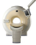
Philips continues to expand the frontiers of utra high field MRI with the introduction of the new Intera Achieva 3.0T™. Its powerful future-safe platform shares all the advantages of the Achieva family and covers applications throughout the whole body.
Device Information and Specification
CLINICAL APPLICATION
Whole body
CONFIGURATION
Short bore compact
Standard: T/R-head, Body, Optional phased array: CTL spine; Optional SENSE coils: Torso, Cardiac, Head, Knee, Breast, Neurovascular, Flex M, Flex L
Optional
SYNCHRONIZATION
ECG/peripheral: Optional/yes, respiratory gating
PULSE SEQUENCES
SE, Modified-SE, IR (T1, T2, PD), STIR, FLAIR, SPIR, FFE, T1-FFE, T2-FFE, Balanced FFE, TFE, Balanced TFE, Dynamic, Keyhole, 3D, Multi Chunk 3D, Multi Stack 3D, K Space Shutter, MTC, TSE, Dual IR, DRIVE, EPI, Cine, 2DMSS, DAVE, Mixed Mode; Angiography: Inflow MRA, TONE, PCA, CE MRA
IMAGING MODES
Single Slice 2D , Multi Single Slice 2D, Multi Slice 2D, 3D, Multi Chunk 3D, Multi Stack 3D
Minimum TR
1.1 msec
FOV
Over 40 cm
0.05 mm
128 x 128, 256 x 256,512 x 512,1024 x 1024 (64 for Bold img)
MEASURING MATRIX
Variable in 1% increments
PIXEL INTENSITY
Lum.: 120 cd/m2; contrast: 150:1
Variable (op. param. depend.)
35 x 35 cm
MAGNET WEIGHT
5500 kg
H*W*D
206 x 200 x 167 cm
POWER REQUIREMENTS
380/400 V
CRYOGEN USE
0.15 L/hr helium
STRENGTH
up to 80 mT/m
5-GAUSS FRINGE FIELD
3.0 m / 5.2 m
Passive and dynamic, 1st order std./2nd opt.
spacer

• View the DATABASE results for 'Intera Achieva 3.0T™' (2).Open this link in a new window

 
Further Reading:
  News & More:
Faster speed, better spatial resolution lead 3T benefits
   by sipi.usc.edu    
MRI Resources 
Intraoperative MRI - Supplies - Quality Advice - Resources - Implant and Prosthesis pool - Manufacturers
 
Lung ImagingMRI Resource Directory:
 - Lung Imaging -
 
Lung imaging is furthermore a challenge in MRI because of the predominance of air within the lungs and associated susceptibility issues as well as low signal to noise of the inflated lung parenchyma. Cardiac and respiratory triggered or breath hold sequences allow diagnostic imaging, however a comparable image quality with computed tomography is still difficult to achieve.
Assumptions for lung MRI:
•
Low signal to noise ratio of the inherently low lung proton density.
•
Cardiac and respiratory motion artifacts.
•
Magnetic susceptibility effects of large magnetic field gradients.
•
Very short transverse relaxation times and significant diffusion yielding short T2 (30-70 msec), short T2* (1-3 msec), and additional long T1 relaxation times (1300-1500 msec).
•
The extreme short T2 values are responsible for a fast signal decay during a single shot readout, resulting in blurring.

The current trends in MRI are the use of new imaging technologies and increasingly powerful magnetic fields. Among these technologies are parallel imaging techniques as well as ventilation agents like hyperpolarized helium for the use as an inert inhalational contrast agent to study lung ventilation properties. With hyperpolarized gases clear images of the lungs can be obtained without using a large magnetic field (see also back projection imaging). Single shot sequences (e.g. TSE or Half Fourier Acquisition Single Shot Turbo Spin Echo HASTE) used in lung MR imaging benefits from parallel imaging techniques due to reduced relaxation time effects during the echo train and therefore reduced image blurring as well as reduced motion artifacts.
In the future, more effective contrast agents may provide an alternative solution to the need for high field MRI. Dynamic contrast enhanced MRI perfusion has demonstrated a potential in the diagnosis of pulmonary embolism or to characterize lung cancer and mediastinal tumors. 3D contrast enhanced magnetic resonance angiography of the thoracic vessel.

See also the related poll result: 'MRI will have replaced 50% of x-ray exams by'
 
Images, Movies, Sliders:
 Anatomic Imaging of the Lungs  Open this link in a new window
      

Courtesy of  Robert R. Edelman
 Normal Lung Gd Perfusion MRI  Open this link in a new window
      

Courtesy of  Robert R. Edelman

 MRI Thorax Basal Plane  Open this link in a new window
 
Radiology-tip.comradLung Scintigraphy
spacer

• View the DATABASE results for 'Lung Imaging' (7).Open this link in a new window


• View the NEWS results for 'Lung Imaging' (3).Open this link in a new window.
 
Further Reading:
  Basics:
A safer approach for diagnostic medical imaging
Monday, 29 September 2014   by www.eurekalert.org    
Parallel Lung Imaging(.pdf)
  News & More:
Chest MRI a viable alternative to chest CT in COVID-19 pneumonia follow-up
Monday, 21 September 2020   by www.healthimaging.com    
CT Imaging Features of 2019 Novel Corona virus (2019-nCoV)
Tuesday, 4 February 2020   by pubs.rsna.org    
Polarean Imaging Phase III Trial Results Point to Potential Improvements in Lung Imaging
Wednesday, 29 January 2020   by www.diagnosticimaging.com    
Low Power MRI Helps Image Lungs, Brings Costs Down
Thursday, 10 October 2019   by www.medgadget.com    
Chest MRI Using Multivane-XD, a Novel T2-Weighted Free Breathing MR Sequence
Thursday, 11 July 2019   by www.sciencedirect.co    
Researchers Review Importance of Non-Invasive Imaging in Diagnosis and Management of PAH
Wednesday, 11 March 2015   by lungdiseasenews.com    
New MRI Approach Reveals Bronchiectasis' Key Features Within the Lung
Thursday, 13 November 2014   by lungdiseasenews.com    
MRI techniques improve pulmonary embolism detection
Monday, 19 March 2012   by medicalxpress.com    
  News & More:
Partnership with VIDA to streamline adoption of advanced MRI of the lungs
Monday, 11 September 2023   by www.itnonline.com    
Searchterm 'High Field MRI' was also found in the following services: 
spacer
News  (36)  Resources  (10)  Forum  (1)  
 
MAGNETOM Spectra
 
www.healthcare.siemens.com/magnetic-resonance-imaging/3t-mri-scanner/magnetom-spectra From Siemens Medical Systems;
Received FDA clearance in 2012.
The MAGNETOM Spectra is a cost-optimized high field MRI system with Tim 4G and Dot technologies. The system consumes less energy compared to other 3 Tesla scanners. The magnet-cooling helium is contained in a closed loop, which prevents the gas from escaping and reduces the need for refills. TimTX includes innovative techniques in the radio frequency excitation hardware as well as new application and processing features enabling uniform RF distribution in all body regions.
Device Information and Specification
CLINICAL APPLICATION
Whole Body
CONFIGURATION
Short bore
3 Tesla
Head, spine, torso/ body coil, neurovascular, neck and multi-purpose flex coils. Peripheral vascular, breast, shoulder, knee, wrist, foot//ankle, endorectal optional.
CHANNELS
24
Chemical shift imaging, single voxel spectroscopy
IMAGING TECHNIQUES
iPAT, mSENSE and GRAPPA (image, k-space), noncontrast angiography, radial motion compensation
FOV
0.5 - 45 cm
BORE DIAMETER
or W x H
At isocenter: L-R 60 cm
TABLE CAPACITY
200 kg
MAGNET WEIGHT
7200 kg
DIMENSION H*W*D (gantry included)
173 x 231 x 219 cm
5-GAUSS FRINGE FIELD
2.6 m / 4.6 m
CRYOGEN USE
Zero boil off rate, approx. 10 years
COOLING SYSTEM
Water; single cryogen, 2 stage refrigeration
up to 125 T/m/s
MAX. AMPLITUDE
33 mT/m
Passive, active; first order standard, second order optional
POWER REQUIREMENTS
380 / 400 / 420 / 440 / 460 / 480 V, 3-phase + ground; connection value with chiller 100 kvA /without chiller 60 kVA
spacer
MRI Resources 
Implant and Prosthesis - Guidance - Contrast Agents - Hospitals - Spectroscopy pool - Spectroscopy
 
MRI SafetyMRI Resource Directory:
 - Safety -
 
There are different types of contraindications that would prevent a person from being examined with an MRI scanner. MRI systems use strong magnetic fields that attract any ferromagnetic objects with enormous force. Caused by the potential risk of heating, produced from the radio frequency pulses during the MRI procedure, metallic objects like wires, foreign bodies and other implants needs to be checked for compatibility. High field MRI requires particular safety precautions. In addition, any device or MRI equipment that enters the magnet room has to be MR compatible. MRI examinations are safe and harmless, if these MRI risks are observed and regulations are followed.

Safety concerns in magnetic resonance imaging include:
•
the magnetic field strength;
•
possible 'missile effects' caused by magnetic forces;
•
the potential for heating of body tissue due to the application of the radio frequency energy;
•
the effects on implanted active devices such as cardiac pacemakers or insulin pumps;
•
magnetic torque effects on indwelling metal (clips, etc.);
•
the audible acoustic noise;
•
danger due to cryogenic liquids;
•
the application of contrast medium;
mri safety guidance
MRI Safety Guidance
It is important to remember when working around a superconducting magnet that the magnetic field is always on. Under usual working conditions the field is never turned off. Attention must be paid to keep all ferromagnetic items at an adequate distance from the magnet. Ferromagnetic objects which came accidentally under the influence of these strong magnets can injure or kill individuals in or nearby the magnet, or can seriously damage every hardware, the magnet itself, the cooling system, etc.. See MRI resources Accidents.
The doors leading to a magnet room should be closed at all times except when entering or exiting the room. Every person working in or entering the magnet room or adjacent rooms with a magnetic field has to be instructed about the dangers. This should include the patient, intensive-care staff, and maintenance-, service- and cleaning personnel, etc..
The 5 Gauss limit defines the 'safe' level of static magnetic field exposure. The value of the absorbed dose is fixed by the authorities to avoid heating of the patient's tissue and is defined by the specific absorption rate. Leads or wires that are used in the magnet bore during imaging procedures, should not form large-radius wire loops. Leg-to-leg and leg-to-arm skin contact should be prevented in order to avoid the risk of burning due to the generation of high current loops if the legs or arms are allowed to touch. The patient's skin should not be in contact with the inner bore of the magnet.
The outflow from cryogens like liquid helium is improbable during normal operation and not a real danger for patients.
The safety of MRI contrast agents is tested in drug trials and they have a high compatibility with very few side effects. The variations of the side effects and possible contraindications are similar to X-ray contrast medium, but very rare. In general, an adverse reaction increases with the quantity of the MRI contrast medium and also with the osmolarity of the compound.

See also 5 Gauss Fringe Field, 5 Gauss Line, Cardiac Risks, Cardiac Stent, dB/dt, Legal Requirements, Low Field MRI, Magnetohydrodynamic Effect, MR Compatibility, MR Guided Interventions, Claustrophobia, MRI Risks and Shielding.
Radiology-tip.comradRadiation Safety,  Ionizing Radiation
spacer
Medical-Ultrasound-Imaging.comUltrasound Safety,  Absorbed Dose
spacer

• View the DATABASE results for 'MRI Safety' (42).Open this link in a new window


• View the NEWS results for 'MRI Safety' (13).Open this link in a new window.
 
Further Reading:
  Basics:
MRI Safety
2001   by www.fda.gov    
What MRI Sequences Produce the Highest Specific Absorption Rate (SAR), and Is There Something We Should Be Doing to Reduce the SAR During Standard Examinations?
Thursday, 16 April 2015   by www.ajronline.org    
Contrast Agents: Safety Profile
   by www.clinical-mri.com    
  News & More:
How safe is 7T MRI for patients with neurosurgical implants?
Thursday, 17 November 2022   by healthimaging.com    
Newer Heart Devices Safe During MRI
Monday, 23 August 2004   by www.hospimedica.com    
Study: Face Masks Unsafe in MRI Machines
Wednesday, 13 July 2022   by www.laboratoryequipment.com    
COVID-19: Attention shifts to MRI infection control
Thursday, 9 July 2020   by https://www.auntminnieeurope.com/index.aspx?sec=ser§sub=def§pag=dis§ItemID=619012    
FDA Releases New Guidance On Establishing Safety, Compatibility Of Passive Implants In MR Environments
Tuesday, 16 December 2014   by www.meddeviceonline.com    
Modern Implantable Heart Devices Safe For Use In MRI Scans
Wednesday, 16 March 2005   by www.sciencedaily.com    
MRI Resources 
Nerve Stimulator - Spectroscopy pool - Open Directory Project - Brain MRI - Lung Imaging - Intraoperative MRI
 
previous      6 - 10 (of 35)     next
Result Pages : [1]  [2 3 4]  [5 6 7]
 Random Page
 
Share This Page
FacebookTwitterLinkedIn

MR-TIP    
Community   
User
Pass
Forgot your UserID/Password ?    


New acceleration techniques will :
reduce scan times 
cause artifacts 
increase expenses 
be useful if you have a lot of experience 
doesn't do much 
never heard of 

Look
      Ups


MR-TIP.com uses cookies! By browsing MR-TIP.com, you agree to our use of cookies.

Magnetic Resonance - Technology Information Portal
Member of SoftWays' Medical Imaging Group - MR-TIP • Radiology-TIP • Medical-Ultrasound-Imaging • 
Copyright © 2003 - 2024 SoftWays. All rights reserved. [ 21 November 2024]
Terms of Use | Privacy Policy | Advertising
 [last update: 2024-02-26 03:41:00]