 | Info
Sheets |
| | | | | | | | | | | | | | | | | | | | | | | | |
 | Out-
side |
| | | | |
|
| | | | |
Result : Searchterm 'High Field MRI' found in 1 term [ ] and 16 definitions [ ] and 16 definitions [ ], (+ 18 Boolean[ ], (+ 18 Boolean[ ] results ] results
| previous 26 - 30 (of 35) nextResult Pages :  [1] [1]  [2 3 4] [2 3 4]  [5 6 7] [5 6 7] |  | |  | Searchterm 'High Field MRI' was also found in the following services: | | | | |
|  |  |
| |
|
Device Information and Specification CLINICAL APPLICATION Whole body Quadrature, solenoid and multi-channel configurations SE, FE, IR, FastSE, FastIR, FastFLAIR, Fast STIR, FastFE, FASE, Hybrid EPI, Multi Shot EPI; Angiography: 2D(gate/non-gate)/3D TOF, SORS-STC IMAGING MODES Single, multislice, volume study POWER REQUIREMENTS 380/400/415/440/480 V COOLING SYSTEM TYPE Cryogenless | |  | | | |
|  | |  |  |  |
| |
|

Swiss-based, formerly Bruker AG - split on the 5th October 2001 into the groups: Bruker Daltonics ( Mass spectroscopy), Bruker Optics (Infrared spectroscopy), Bruker AXS (X-ray spectroscopy) and Bruker BioSpin (The largest part, the NMR business core, the EPR and the Tomography activities).
Product Lines:
•
PharmaScan® - MRI//MRS systems tailored to high-throughput and routine applications in pharmaceutical research.
Product Specification
Contact Information
Please see Bruker BioSpin AG's
| |  | |
• View the NEWS results for 'Bruker BioSpin AG' (1).
| | | | |  Further Reading: Further Reading: | News & More:
|
|
| |
|  | |  |  |  |
| |
|

The company is a member of the Bracco Group, a highly innovative healthcare group and world leader in global integrated solutions for the diagnostic medical imaging field. The Bracco Group is headquartered in Milan, Italy. Its North American operations consist of Bracco Diagnostics and Bracco Research USA, both located in Princeton, New Jersey. Bracco Diagnostics is one of the fastest growing developers and marketers of diagnostic pharmaceuticals in North America, with products for various imaging applications, including Isovue® (iopamidol - X-ray contrast agent), ProHance® ( gadoteridol - MRI contrast agent), SonoVue® ( ultrasound contrast agent) and nuclear medicine products.
Gadoteridol has been available in Europe and the USA for several years.
Holder of the Marketing Authorization:
Bracco International B.V. - Strawinskylaan 3051 - 1077 ZX Amsterdam
The Netherlands. (Contact: Kirk Deeter, Phone: +NL-303-838-8708)
MRI Contrast Agents:
Contact Information
Please see Bracco Diagnostics, Inc.'s
| |  | |
• View the DATABASE results for 'Bracco Diagnostics, Inc.' (2).
| | | | |  Further Reading: Further Reading: | News & More:
|
|
| |
|  |  | Searchterm 'High Field MRI' was also found in the following services: | | | | |
|  |  |
| |
|

(EPI) Echo planar imaging is one of the early magnetic resonance imaging sequences (also known as Intascan), used in applications like diffusion, perfusion, and functional magnetic resonance imaging. Other sequences acquire one k-space line at each phase encoding step. When the echo planar imaging acquisition strategy is used, the complete image is formed from a single data sample (all k-space lines are measured in one repetition time) of a gradient echo or spin echo sequence (see single shot technique) with an acquisition time of about 20 to 100 ms.
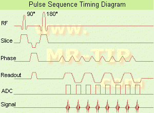
The pulse sequence timing diagram illustrates an echo planar imaging sequence from spin echo type with eight echo train pulses. (See also Pulse Sequence Timing Diagram, for a description of the components.)
In case of a gradient echo based EPI sequence the initial part is very similar to a standard gradient echo sequence. By periodically fast reversing the readout or frequency encoding gradient, a train of echoes is generated.
EPI requires higher performance from the MRI scanner like much larger gradient amplitudes. The scan time is dependent on the spatial resolution required, the strength of the applied gradient fields and the time the machine needs to ramp the gradients.
In EPI, there is water fat shift in the phase encoding direction due to phase accumulations. To minimize water fat shift (WFS) in the phase direction fat suppression and a wide bandwidth (BW) are selected. On a typical EPI sequence, there is virtually no time at all for the flat top of the gradient waveform. The problem is solved by "ramp sampling" through most of the rise and fall time to improve image resolution.
The benefits of the fast imaging time are not without cost. EPI is relatively demanding on the scanner hardware, in particular on gradient strengths, gradient switching times, and receiver bandwidth. In addition, EPI is extremely sensitive to image artifacts and distortions. | |  | |
• View the DATABASE results for 'Echo Planar Imaging' (19).
| | |
• View the NEWS results for 'Echo Planar Imaging' (1).
| | | | |  Further Reading: Further Reading: | Basics:
|
|
| |
|  | |  |  |  |
| |
|

From Esaote S.p.A.;
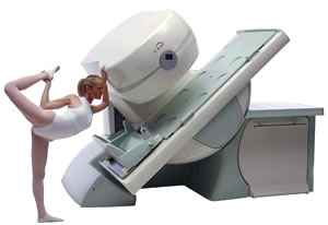
Esaote introduced the new G-SCAN at the RSNA in Dec. 2004. The G-SCAN covers almost all musculoskeletal applications including the spine. The tilting gantry is designed for scanning in weight-bearing positions. This unique MRI scanner is developed in line with the Esaote philosophy of creating high quality MRI systems that are easy to install and that have a low breakeven point.
Device Information and Specification
SE, GE, IR, STIR, TSE, 3D CE, GE-STIR, 3D GE, ME, TME, HSE
100 up to 350 mm, 25 mm displayed
POWER REQUIREMENTS
100/110/200/220/230/240 V
| |  | |
• View the DATABASE results for 'G-SCAN' (3).
| | | | |
|  | |  |  |
|  | |
|  | | |
|
| |
 | Look
Ups |
| |