Magnetic Resonance - Technology Information Portal Welcome to MRI Technology
Info
  Sheets

Out-
      side


 'Hybrid Spectroscopy' 
SEARCH FOR    
 
  2 3 5 A B C D E F G H I J K L M N O P Q R S T U V W X Y Z
Result : Searchterm 'Hybrid Spectroscopy' found in 1 term [] and 0 definition [], (+ 6 Boolean[] results
1 - 5 (of 7)     next
Result Pages : [1]  [2]
MRI Resources 
Jobs - Brain MRI - RIS - MRCP - - Societies
 
Hybrid Spectroscopy
 
The combination of single volume spectroscopy and chemical shift imaging. The chemical shift measurement is performed over a selectively-excited volume of interest. Areas with strong distorting signal are not stimulated and therefore do not append signal to the spectra.

See also Hybrid Imaging.
spacer
 
• Share the entry 'Hybrid Spectroscopy':  Facebook  Twitter  LinkedIn  
 
Further Reading:
  News & More:
Near-IR Spectroscopy Performs Challenging Breast Imaging
Tuesday, 18 February 2014   by www.novuslight.com    
MRI Resources 
Calculation - Breast Implant - MRI Accidents - Equipment - Process Analysis - Fluorescence
 
Excelart AG™ with PianissimoInfoSheet: - Devices -
Intro, 
Types of Magnets, 
Overview, 
etc.MRI Resource Directory:
 - Devices -
 
http://www.medical.toshiba.com/clinical/radiology/15texcelart.htm From Toshiba America Medical Systems Inc.;
the EXCELART is a superconducting whole body MRI system with a short wide-bore magnet, operating at 1.5 T. It features powerful high-speed gradients with a revolutionary gradient acoustic noise reduction system: Pianissimo. The dramatic reduction of gradient acoustic noise by Pianissimo greatly enhances patient comfort during exams. The standard array platform and a wide range of array coils ensure excellent images. A powerful 64-bit RISC-based computer system and newly developed array processor realize high productivity.
Device Information and Specification
CLINICAL APPLICATION
Whole body
CONFIGURATION
Cylindrical Wide Short Bore
Opt. (WIP) Single and Multi Voxel
SYNCHRONIZATION
ECG: standard; peripheral: optional, respiratory gating
PULSE SEQUENCES
SE, FE, IR, FastSE, FastIR, FastFLAIR, Fast STIR, FastFE, FASE, Hybrid EPI, Multi Shot EPI; Angiography: 2D(gate/non-gate)/3D TOF, SORS-STC
IMAGING MODES
Single, multislice, volume study
TR
3.5-30000 msec
TE
8 msec min. SE; 1.2 msec min. FE
SINGLE/MULTI SLICE
less than 0.015 (256x256)
FOV
2 cm
1.0 min. 2-DFT: 0.2 min. 3-DFT
Up to 1024
MEASURING MATRIX
32-1024, phase;; 64-1024, freq.
PIXEL INTENSITY
256 gray levels
BORE DIAMETER
or W x H
65.5 cm, patient aperture
MAGNET WEIGHT
4050 kg (bare magnet incl. L-He)
H*W*D
235 x 219 x 199 cm
COOLING SYSTEM TYPE
Closed-loop water-cooled
CRYOGEN USE
Liquid helium: approx. less than 0.05 L/hr
STRENGTH
25 mT/m
5-GAUSS FRINGE FIELD
2.5 m / 4.0 m
Passive, active, auto-active
spacer

• View the DATABASE results for 'Excelart AG™ with Pianissimo' (2).Open this link in a new window

MRI Resources 
Libraries - Abdominal Imaging - Bioinformatics - MRI Reimbursement - Calculation - Blood Flow Imaging
 
Excelart XG™ with PianissimoInfoSheet: - Devices -
Intro, 
Types of Magnets, 
Overview, 
etc.MRI Resource Directory:
 - Devices -
 
http://www.medical.toshiba.com/clinical/radiology/15texcelart.htm From Toshiba America Medical Systems Inc.;
the EXCELART is a superconducting whole body MRI system with a short wide-bore magnet, operating at 1.5 T. It features powerful high-speed gradients with a revolutionary gradient acoustic noise reduction system: Pianissimo. The dramatic reduction of gradient acoustic noise by Pianissimo greatly enhances patient comfort during exams. The standard array platform and a wide range of array coils ensure excellent images. A powerful 64-bit RISC-based computer system and newly developed array processor realize high productivity.
Device Information and Specification
CLINICAL APPLICATION
Whole body
CONFIGURATION
Cylindrical Wide Short Bore
Optional (WIP)
SYNCHRONIZATION
ECG/peripheral, respiratory gating
PULSE SEQUENCES
SE, FE, IR, FastSE, FastIR, FastFLAIR, Fast STIR, FastFE, FASE, Hybrid EPI, Multi Shot EPI; Angiography: 2D(gate/non-gate)/3D TOF, SORS-STC
IMAGING MODES
Single, multislice, volume study
TR
2.6-30000 msec
TE
8 msec min. SE; 0.9 msec min. FE
SINGLE/MULTI SLICE
less than 0.011 (256x256)
FOV
2 cm
1.0 min. 2-DFT: 0.2 min. 3-DFT
Up to 1024
MEASURING MATRIX
32-1024, phase;; 64-1024, freq.
PIXEL INTENSITY
256 gray levels
BORE DIAMETER
or W x H
65.5 cm, patient aperture
MAGNET WEIGHT
4050 kg (bare magnet incl. L-He)
H*W*D
235 x 219 x 199 cm
POWER REQUIREMENTS
380/400/415/440/480 V
COOLING SYSTEM TYPE
Closed-loop water-cooled
CRYOGEN USE
Liquid helium: approx. less than 0.05 L/hr
STRENGTH
25 mT/m
5-GAUSS FRINGE FIELD
2.5 m / 4.0 m
Passive, active, auto-active
spacer

• View the DATABASE results for 'Excelart XG™ with Pianissimo' (2).Open this link in a new window

 
Further Reading:
  News & More:
Angio, cardiac imaging top list for MR and CT
1999   by www.diagnosticimaging.com    
MRI Resources 
Hospitals - Contrast Agents - Artifacts - Homepages - RIS - Image Quality
 
OPART™InfoSheet: - Devices -
Intro, 
Types of Magnets, 
Overview, 
etc.MRI Resource Directory:
 - Devices -
 
www3.toshiba.co.jp/medical/products/mr/opart/index.htm From Toshiba America Medical Systems Inc.;
OPART™ is a 0.35 T superconducting open MR system featuring cryogenless operation. Superconducting speed and performance are combined with the patient access advantages of open MRI design for unmatched clinical versatility. OPART™ features innovative technologies such as digital RF, high speed gradients and optimized RF coils, which support advanced MRI applications.
Device Information and Specification
CLINICAL APPLICATION
Whole body
CONFIGURATION
Quadrature, solenoid and multi-channel configurations
SYNCHRONIZATION
ECG/peripheral optional, respiratory gating
PULSE SEQUENCES
SE, FE, IR, FastSE, FastIR, FastFLAIR, Fast STIR, FastFE, FASE, Hybrid EPI, Multi Shot EPI; Angiography: 2D(gate/non-gate)/3D TOF, SORS-STC
IMAGING MODES
Single, multislice, volume study
55 cm, vertical opening
POWER REQUIREMENTS
380/400/415/440/480 V
COOLING SYSTEM TYPE
Cryogenless
STRENGTH
25 mT/m
Passive, active
spacer

• View the DATABASE results for 'OPART™' (2).Open this link in a new window

MRI Resources 
Safety Products - Spectroscopy pool - Pediatric and Fetal MRI - Fluorescence - MRI Centers - Most Wanted
 
Biograph mMR
 
www.healthcare.siemens.com/magnetic-resonance-imaging/mr-pet-scanner/biograph-mmr FDA cleared and CE Mark 2011.
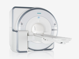
The Biograph mMR has a fully-integrated design for simultaneous PET/MRI imaging. The dedicated hardware includes solid-state, avalanche photodiode PET detector and adapted, PET-compatible MR coils.
The possibility of truly simultaneous operation allows the acquisition of several magnetic resonance imaging (MRI) sequences during the positron emission tomography (PET) scan, without increasing the examination time.
See also Hybrid Imaging.
Device Information and Specification
CLINICAL APPLICATION
Whole Body
CONFIGURATION
Simultaneous PET/MRI
3 Tesla
Full range of MRI coils
CHANNELS
up to 32
PET ACQUISITION MODES
3D
MRI ACQUISITION MODES
2D/3D, spectroscopy;; iPAT, GRAPPA (k-space), noncontrast angiography, radial motion compensation
PET EFFECTIVE AXIAL FOV
26 cm (typical overlap 23%)
MRI FOV
A-P 45, R-L 50, H-F 50 cm
PET RING DIAMETER
65.6 cm
PATIENT SCAN RANGE
199 cm
HORIZONTAL SPEED
200 mmsec
TABLE CAPACITY
200 kg
PET DETECTOR
Solid state, 4032 avalanche photo diodes
DETECTOR SCINTILLATION MATERIAL
LSO, 28672 crystals
CRYSTAL SIZE
4 x 4 x 20 mm
MAGNET WEIGHT
9000 kg
DIMENSION H*W*D (gantry included)
335 x 230 x 242 cm (finshed covers)
5-GAUSS FRINGE FIELD
2.6 m / 4.6 m
CRYOGEN USE
Zero boil off rate, approx. 10 years
COOLING SYSTEM
PET system: water; MRI system: water
up to 200 T/m/s
MAX. AMPLITUDE
45 mT/m
Aautomatic, patient specific shim; active shim 3 linear and 5 non-linear channels (seond order)
POWER REQUIREMENTS
380 / 400 / 420 / 440 / 460 / 480 V, 3-phase + ground; Total system 110kW
spacer
 
Further Reading:
  Basics:
Performance Measurements of the Siemens mMR Integrated Whole-Body PET/MR Scanner
Friday, 11 November 2011   by jnm.snmjournals.org    
Global Trends in Hybrid Imaging
Monday, 1 November 2010   by pubs.rsna.org    
  News & More:
Positron Emission Tomographic Imaging in Stroke
Monday, 28 December 2015   by www.ncbi.nlm.nih.gov    
A world's first in imaging – integrated whole-body molecular MR system available for clinical use testing
Thursday, 18 November 2010   by www.siemens.com    
FDA Clears New System to Perform Simultaneous PET, MRI Scans
Friday, 10 June 2011   by www.prnewswire.com    
Frost & Sullivan Award Underlines Siemens Healthcare's Contribution to New Product Innovation in the North American Medical Imaging Market
Wednesday, 13 July 2011   by multivu.prnewswire.com    
MRI Resources 
Image Quality - MRA - Raman Spectroscopy - Journals - Case Studies - MRI Technician and Technologist Career
 
     1 - 5 (of 7)     next
Result Pages : [1]  [2]
 Random Page
 
Share This Page
FacebookTwitterLinkedIn

MR-TIP    
Community   
User
Pass
Forgot your UserID/Password ?    


MRI is trending to low field magnets :
reduced costs will lead to this change 
AI will close the gap to high field 
only in remote areas 
is only temporary 
never 

Look
      Ups


MR-TIP.com uses cookies! By browsing MR-TIP.com, you agree to our use of cookies.

Magnetic Resonance - Technology Information Portal
Member of SoftWays' Medical Imaging Group - MR-TIP • Radiology-TIP • Medical-Ultrasound-Imaging • 
Copyright © 2003 - 2024 SoftWays. All rights reserved. [ 21 November 2024]
Terms of Use | Privacy Policy | Advertising
 [last update: 2024-02-26 03:41:00]