Magnetic Resonance - Technology Information Portal Welcome to MRI Technology
Info
  Sheets

Out-
      side


 'Imaging' 
SEARCH FOR    
 
  2 3 5 A B C D E F G H I J K L M N O P Q R S T U V W X Y Z
Result : Searchterm 'Imaging' found in 72 terms [] and 403 definitions []
previous     16 - 20 (of 475)     next
Result Pages : [1 2 3 4 5 6 7 8 9 10 11 12 13 14 15]  [16 17 18 19 20 ... ]
Searchterm 'Imaging' was also found in the following services: 
spacer
News  (1520)  Resources  (268)  Forum  (88)  
 
Blood Flow ImagingMRI Resource Directory:
 - Blood Flow Imaging -
 
MR imaging techniques capable to provide maps of cerebral activity. All these techniques are based on indirect assessment of local cerebral haemodynamics that have been demonstrated to be closely related to cerebral activity.
Two kinds of techniques have been developed:
based on the assessment of the decrease in the content of deoxyhaemoglobin in local activated tissue that can be revealed as an increase of signal on T2* and T2 weighted sequences in which deoxyhaemoglobin has low signal (see Blood Oxygenation Level Dependent Contrast)
based on the time of flight or flow-related enhancement that is revealed either directly with T1 weighted images or through the use of modified angiographic bolus tracking techniques.
 
Images, Movies, Sliders:
 Circle of Willis, Time of Flight, MIP  Open this link in a new window
    
SlidersSliders Overview

 
spacer
 
• Related Searches:
    • Functional Magnetic Resonance Imaging
    • Brain MRI
    • Velocity Encoding
    • Arterial Spin Labeling
    • Magnetic Resonance Angiography MRA
 
Further Reading:
  News & More:
Turbo-FLASH Based Arterial Spin Labeled Perfusion MRI at 7 T
Thursday, 20 June 2013   by www.plosone.org    
Non-invasive MRI technique distinguishes between Alzheimer's and frontotemporal dementia
Saturday, 18 June 2005   by www.eurekalert.org    
MRI Resources 
Quality Advice - Pathology - Abdominal Imaging - Diffusion Weighted Imaging - Libraries - Most Wanted
 
Multi Echo Imaging
 
Multi echo imaging sequences use a series of echoes acquired as a train following after a single excitation pulse. Multiple symmetrical or asymmetrical echoes can be acquired, typically T2 weighted. In spin echo imaging, each echo is formed by a 180° pulse, but also a FSE (TSE, RARE) or EPI sequence can be used. As a difference to a normal fast spin echo sequence, in multi echo imaging, separate images are produced from each echo of the train with different T2 weightings. The signal height reduces with transverse relaxation. This drop in signal can be used to calculate a pure T2 image.

See also Fast Spin Echo.
spacer

• View the DATABASE results for 'Multi Echo Imaging' (3).Open this link in a new window

 
Further Reading:
  Basics:
A very simple, robust and fast method for estimating and displaying average time constants of T2 decays from multiecho MRI images using color intensity projections
   by arxiv.org    
What MRI Sequences Produce the Highest Specific Absorption Rate (SAR), and Is There Something We Should Be Doing to Reduce the SAR During Standard Examinations?
Thursday, 16 April 2015   by www.ajronline.org    
  News & More:
Automatic Mapping Extraction from Multiecho T2-Star Weighted Magnetic Resonance Images for Improving Morphological Evaluations in Human Brain
Wednesday, 5 June 2013   by www.hindawi.com    
RARE
Monday, 3 December 2012   by www2.warwick.ac.uk    
MRI Resources 
Veterinary MRI - Online Books - Pediatric and Fetal MRI - Liver Imaging - Functional MRI - Stimulator pool
 
Volumetric Imaging
 
Volumetric imaging is a 3D technique where all the MRI signals are collected from the entire tissue sample and imaged as a whole entity, therefore providing a high signal to noise ratio. The acquisition of isotropic voxels or thin slices with high spatial resolution allows to create multiplanar reconstructions in all planes; a compensation for the usually longer scan time. The acquisition time can be reduced by parallel imaging technique.
New T2 weighted variants of 3D sequences (FSE-XETA, T2-SPACE, VISTA) have been introduced that differ from conventional FSE sequences. An echo train containing up to 200 echoes obtained at a minimum echo spacing allows very fast acquisition. A flip angle modulation (flip angle sweep - FAS) during the FSE readout carries magnetization as long as possible to avoid blurring and provide optimal signal at the effective echo time. This type of imaging is well suited for brain and spine MRI procedures.
Newer T1 weighted variants include Liver Acquisition with Volume Acquisition (LAVA) and T1W High Resolution Isotropic Volume Examination (THRIVE), which have advantages for dynamic breath hold imaging in liver and abdominal examinations.

See also Volume Imaging, 3 Dimensional Imaging.
 
Images, Movies, Sliders:
 Brain MRI Sagittal T1 001  Open this link in a new window
    
 Circle of Willis, Time of Flight, MIP  Open this link in a new window
    
SlidersSliders Overview

 MRI of the Skull Base  Open this link in a new window
    
SlidersSliders Overview

 
spacer

• View the DATABASE results for 'Volumetric Imaging' (4).Open this link in a new window


• View the NEWS results for 'Volumetric Imaging' (1).Open this link in a new window.
 
Further Reading:
  Basics:
Cutting Edge Imaging of THE Spine
February 2007   by www.pubmedcentral.nih.gov    
3-D VOLUMETRIC IMAGING FOR STEREOTACTIC LESIONAL AND DEEP BRAIN STIMULATION SURGERY
Searchterm 'Imaging' was also found in the following services: 
spacer
News  (1520)  Resources  (268)  Forum  (88)  
 
Breath Hold ImagingMRI Resource Directory:
 - Abdominal Imaging -
 
Breath hold imaging in MRI is a technique with one ore more stoppage of breathing during the sequence and require therefore a short scan time. Breath hold techniques are used with fast gradient echo sequences in thoracic or abdominal regions with much respiratory movement.
Breath hold cine MRI techniques are used in cardiovascular imaging and provide detailed views of the beating heart in different cardiac axes.
Breath hold imaging requires the full cooperation of the patient, caused by usual MRI scan times from 15 to 20 sec.. In some cases breath holding can be practiced outside the MRI scanner to improve patient cooperation with the examination. Shorter scan times e.g. by parallel imaging techniques, or the administration of oxygen can help the patient to hold the breath during the scan.
See also Abdominal Imaging.
 
Images, Movies, Sliders:
 MRI Upper Abdomen T1 with Contrast  Open this link in a new window
 Normal Dual Inversion Fast Spin-echo  Open this link in a new window
 Anatomic Imaging of the Lungs  Open this link in a new window
 
spacer

• View the DATABASE results for 'Breath Hold Imaging' (7).Open this link in a new window

 
Further Reading:
  News & More:
The Effects of Breathing Motion on DCE-MRI Images: Phantom Studies Simulating Respiratory Motion to Compare CAIPIRINHA-VIBE, Radial-VIBE, and Conventional VIBE
Tuesday, 7 February 2017   by www.kjronline.org    
Controlling patient's breathing makes cardiac MRI more accurate
Friday, 13 May 2016   by www.upi.com    
Accurate T1 Quantification Using a Breath-hold Inversion Recovery TrueFISP Sequence
2003   by rsna2003.rsna.org    
MRI Resources 
Non-English - Distributors - General - Liver Imaging - Directories - Homepages
 
Echo Planar ImagingInfoSheet: - Sequences - 
Intro, 
Overview, 
Types of, 
etc.MRI Resource Directory:
 - Sequences -
 
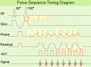
Echo Planar Imaging Timing Diagram (EPI) Echo planar imaging is one of the early magnetic resonance imaging sequences (also known as Intascan), used in applications like diffusion, perfusion, and functional magnetic resonance imaging. Other sequences acquire one k-space line at each phase encoding step. When the echo planar imaging acquisition strategy is used, the complete image is formed from a single data sample (all k-space lines are measured in one repetition time) of a gradient echo or spin echo sequence (see single shot technique) with an acquisition time of about 20 to 100 ms. The pulse sequence timing diagram illustrates an echo planar imaging sequence from spin echo type with eight echo train pulses. (See also Pulse Sequence Timing Diagram, for a description of the components.)
In case of a gradient echo based EPI sequence the initial part is very similar to a standard gradient echo sequence. By periodically fast reversing the readout or frequency encoding gradient, a train of echoes is generated.
EPI requires higher performance from the MRI scanner like much larger gradient amplitudes. The scan time is dependent on the spatial resolution required, the strength of the applied gradient fields and the time the machine needs to ramp the gradients.
In EPI, there is water fat shift in the phase encoding direction due to phase accumulations. To minimize water fat shift (WFS) in the phase direction fat suppression and a wide bandwidth (BW) are selected. On a typical EPI sequence, there is virtually no time at all for the flat top of the gradient waveform. The problem is solved by "ramp sampling" through most of the rise and fall time to improve image resolution.
The benefits of the fast imaging time are not without cost. EPI is relatively demanding on the scanner hardware, in particular on gradient strengths, gradient switching times, and receiver bandwidth. In addition, EPI is extremely sensitive to image artifacts and distortions.
spacer

• View the DATABASE results for 'Echo Planar Imaging' (19).Open this link in a new window


• View the NEWS results for 'Echo Planar Imaging' (1).Open this link in a new window.
 
Further Reading:
  Basics:
New Imaging Method Makes Brain Scans 7 Times Faster
Sunday, 9 January 2011   by www.dailytech.com    
MRI Resources 
Hospitals - Safety Products - Mobile MRI Rental - Brain MRI - Jobs pool - Pregnancy
 
previous      16 - 20 (of 475)     next
Result Pages : [1 2 3 4 5 6 7 8 9 10 11 12 13 14 15]  [16 17 18 19 20 ... ]
 Random Page
 
Share This Page
FacebookTwitterLinkedIn

MR-TIP    
Community   
User
Pass
Forgot your UserID/Password ?    


Next big thing in MRI will be :
AI 
remote operator 
personalized protocols 
helium-free 
molecular MRI 
portable MRI 

Look
      Ups


MR-TIP.com uses cookies! By browsing MR-TIP.com, you agree to our use of cookies.

Magnetic Resonance - Technology Information Portal
Member of SoftWays' Medical Imaging Group - MR-TIP • Radiology-TIP • Medical-Ultrasound-Imaging • 
Copyright © 2003 - 2024 SoftWays. All rights reserved. [ 4 December 2024]
Terms of Use | Privacy Policy | Advertising
 [last update: 2024-02-26 03:41:00]