 | Info
Sheets |
| | | | | | | | | | | | | | | | | | | | | | | | |
 | Out-
side |
| | | | |
|
| | | | |
Result : Searchterm 'Imaging Coil' found in 1 term [ ] and 7 definitions [ ] and 7 definitions [ ], (+ 19 Boolean[ ], (+ 19 Boolean[ ] results ] results
| | 1 - 5 (of 27) nextResult Pages : [1] [2] [3 4 5 6] |  | |  | Searchterm 'Imaging Coil' was also found in the following services: | | | | |
| |  |
| Imaging Coil |   |
| |
|
| | | | | | • Share the entry 'Imaging Coil':    | | |
• View the NEWS results for 'Imaging Coil' (9).
| | | | |  Further Reading: Further Reading: | Basics:
|
|
| |
| | | | |  |
| |
|
General MRI of the abdomen can consist of T1 or T2 weighted spin echo, fast spin echo ( FSE, TSE) or gradient echo sequences with fat suppression and contrast enhanced MRI techniques. The examined organs include liver, pancreas, spleen, kidneys, adrenals as well as parts of the stomach and intestine (see also gastrointestinal imaging). Respiratory compensation and breath hold imaging is mandatory for a good image quality.
T1 weighted sequences are more sensitive for lesion detection than T2 weighted sequences at 0.5 T, while higher field strengths (greater than 1.0 T), T2 weighted and spoiled gradient echo sequences are used for focal lesion detection.
Gradient echo in phase T1 breath hold can be performed as a dynamic series with the ability to visualize the blood distribution. Phases of contrast enhancement include the capillary or arterial dominant phase for demonstrating hypervascular lesions, in liver imaging the portal venous phase demonstrates the maximum difference between the liver and hypovascular lesions, while the equilibrium phase demonstrates interstitial disbursement for edematous and malignant tissues.
Out of phase gradient echo imaging for the abdomen is a lipid-type tissue sensitive sequence and is useful for the visualization of focal hepatic lesions, fatty liver (see also Dixon), hemochromatosis, adrenal lesions and renal masses.
The standards for abdominal MRI vary according to clinical sites based on sequence availability and MRI equipment.
Specific abdominal imaging coils and liver-specific contrast agents targeted to the healthy liver tissue improve the detection and localization of lesions.
See also Hepatobiliary Contrast Agents, Reticuloendothelial Contrast Agents, and Oral Contrast Agents.
For Ultrasound Imaging (USI) see Abdominal Ultrasound at Medical-Ultrasound-Imaging.com. | | | | | |
• View the DATABASE results for 'Abdominal Imaging' (11).
| | |
• View the NEWS results for 'Abdominal Imaging' (3).
| | | | | | Further Reading: | | Basics:
|
|
News & More:
|  |
Assessment of Female Pelvic Pathologies: A Cross-Sectional Study Among Patients Undergoing Magnetic Resonance Imaging for Pelvic Assessment at the Maternity and Children Hospital, Qassim Region, Saudi Arabia
Saturday, 7 October 2023 by www.cureus.com | | |
Higher Visceral, Subcutaneous Fat Levels Predict Brain Volume Loss in Midlife
Wednesday, 4 October 2023 by www.neurologyadvisor.com | | |
Deep Learning Helps Provide Accurate Kidney Volume Measurements
Tuesday, 27 September 2022 by www.rsna.org | | |
CT, MRI for pediatric pancreatitis interobserver agreement with INSPPIRE
Friday, 11 March 2022 by www.eurekalert.org | | |
Clinical trial: Using MRI for prostate cancer diagnosis equals or beats current standard
Thursday, 4 February 2021 by www.eurekalert.org | | |
Computer-aided detection and diagnosis for prostate cancer based on mono and multi-parametric MRI: A review - Abstract
Tuesday, 28 April 2015 by urotoday.com | | |
Nottingham scientists exploit MRI technology to assist in the treatment of IBS
Thursday, 9 January 2014 by www.news-medical.net | | |
New MR sequence helps radiologists more accurately evaluate abnormalities of the uterus and ovaries
Thursday, 23 April 2009 by www.eurekalert.org | | |
MRI identifies 'hidden' fat that puts adolescents at risk for disease
Tuesday, 27 February 2007 by www.eurekalert.org |
|
| |
| | | | | |
| |
|
A RF coil, often a transmit receive coil with a number of wires running along the z-direction, arranged to give a cosine current variation around the circumference of the coil, which looks like a bird cage.
The bird cage coil works on a different principle to conventionally tuned local and surround coils in that it behaves like a tuned transmission line with one complete cycle of standing wave around the circumference. The frequency supply is generated by an oscillator, which is modulated to form a shaped pulse by a product detector controlled by the waveform generator. The signal must be amplified to 1000's of watts. This can be done using either solid state electronics, valves or a combination of both.
The bird cage coil design provides the best field homogeneity of all RF imaging coils.
One advantage is that it is simple to produce an exceedingly uniform B1 radio frequency field over most of the coil's volume, with the result of images with a high degree of uniformity.
A second advantage is that nodes with zero voltage occur 90° away from the driven part of the coil, thus facilitating the introduction of a second signal in quadrature, which produces a circularly polarized radio frequency field.
This type of volume coil is used for brain (head) MRI, or MR imaging of joints, such as the wrist or knees.
See also the related poll result: ' 3rd party coils are better than the original manufacturer coils' | | | | | |
• View the DATABASE results for 'Bird Cage Coil' (4).
| | | | | | Further Reading: | Basics:
|
|
News & More:
| |
| |
| | | Searchterm 'Imaging Coil' was also found in the following services: | | | | |
| | |
| |
|
A coil is a large inductor with a considerable dimension and a defined wavelength, commonly used in configurations for MR imaging. The frequency of the radio frequency coil is defined by the Larmor relationship. The MRI image quality depends on the signal to noise ratio (SNR) of the acquired signal from the patient. Several MR imaging coils are necessary to handle the diversity of applications. Large coils have a large measurement field, but low signal intensity and vice versa (see also coil diameter). The closer the coil to the object, the stronger the signal - the smaller the volume, the higher the SNR. SNR is very important in obtaining clear images of the human body. The shape of the coil depends on the image sampling. The best available homogeneity can be reached by choice of the appropriate coil type and correct coil positioning. Orientation is critical to the sensitivity of the RF coil and therefore the coil should be perpendicular to the static magnetic field.
RF coils can be differentiated by there function into three general categories:
The RF signal is in the range of 10 to 100 MHz. During a typical set of clinical image measurements, the entire frequency spectrum of interest is of the order 10 kHz, which is an extremely narrow band, considering that the center frequency is about 100 MHz. This allows the use of single-frequency matching techniques for coils because their inherent bandwidth always exceeds the image bandwidth. The multi turn solenoid, bird cage coil, single turn solenoid, and saddle coil are typically operated as the transmitter and receiver of RF energy. The surface and phased array coils are typically operated as a receive only coil.
See also the related poll result: ' 3rd party coils are better than the original manufacturer coils' | | | | | |
• View the DATABASE results for 'Radio Frequency Coil' (9).
| | | | | | Further Reading: | Basics:
|
|
News & More:
| |
| |
| | | | | |
| |
|
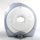
From GE Healthcare;
GE Healthcare has added the Signa HDe 1.5T™, a compact MRI device at an affordable price to its family of MRI products. It has a single electronic cabinet that can be positioned inside the scanner room rather than
in a separate equipment room. The Signa HDe 1.5T can be installed in the same physical location as 0.5T MRI systems with minimal construction costs. According to GE, the installation has been simplified to last only 7 days and has a 30 percent smaller footprint than a typical 1.5T system.
The 1.5T Signa™ HDe MRI system is substantially equivalent to the currently marketed GE 1.5T machines. The data acquisition system supports 1, 4, 8 independent receive channels and multiple independent coil elements per channel during a single acquisition series. The gradient specifications of HDe are lower than other GE Signa 1.5T MRI systems, but it can support clinical applications in cardiac and spectroscopy imaging.
Device Information and Specification CLINICAL APPLICATION Whole body CONFIGURATION Compact short bore 2D 0.7 mm to 20 mm; 3D 0.1 mm to 5 mm 128x512 steps 32 phase encode POWER REQUIREMENTS 480 or 380/415 less than 0.03 L/hr liquid helium | | | |
• View the NEWS results for 'Signa HDe 1.5T™' (1).
| | | | | | Further Reading: | Basics:
|
|
| |
| | | | |
| | | 1 - 5 (of 27) nextResult Pages : [1] [2] [3 4 5 6] |
| |
|
| |
 | Look
Ups |
| |


