 | Info
Sheets |
| | | | | | | | | | | | | | | | | | | | | | | | |
 | Out-
side |
| | | | |
|
| | | | |
Result : Searchterm 'No Phase Wrap' found in 1 term [ ] and 5 definitions [ ] and 5 definitions [ ], (+ 2 Boolean[ ], (+ 2 Boolean[ ] results ] results
| 1 - 5 (of 8) nextResult Pages :  [1] [1]  [2] [2] |  | | |  |  |  |
| |
|
(NPW / PNW - Phase No Wrap) If the receiving RF coil is sensitive to tissue signal arising from outside the desired FOV, this undesired signal may be incorrectly mapped, or wrapped back to a location within the image and is seen as artifact. This problem occurs in the phase encoding direction, where the phases of signal-bearing tissues outside of the FOV in the y-direction are a replication of the phases that are encoded within the FOV.
A user-selectable parameter maps this signal to its correct location outside the FOV, then discards any signal from outside the FOV before displaying the image. No phase wrap works by filling k-space to the same extent, using twice as many phase encoding steps. In order to be able to choose this parameter, in most cases more than an average is necessary.
See Foldover Suppression and Oversampling. | |  | | | | • Share the entry 'No Phase Wrap':    | | | | | | | | | |
|  | |  |  |  |
| |
|
Quick Overview
Please note that there are different common names for this MRI artifact.
DESCRIPTION
Image wrap around
Aliasing is an artifact that occurs in MR images when the scanned body part is larger than field of view ( FOV). As a consequence of the acquired k-space frequencies not being sampled densely enough, whereby portions of the object outside of the desired FOV get mapped to an incorrect location inside the FOV. The cyclical property of the Fourier transform fills the missing data of the right side with data from behind the FOV of the left side and vice versa. This is caused by a too small number of samples acquired in, e.g. the frequency encoding direction, therefore the spectrums will overlap, resulting in a replication of the object in the x direction.
Aliasing in the frequency direction can be eliminated by twice as fast sampling of the signal or by applying frequency specific filters to the received signal.
A similar problem occurs in the phase encoding direction, where the phases of signal-bearing tissues outside of the FOV in the y-direction are a replication of the phases that are encoded within the FOV. Phase encoding gradients are scaled for the field of view only, therefore tissues outside the FOV do not get properly phase encoded relative to their actual position and 'wraps' into the opposite side of the image.

Image Guidance
| |  | |
• View the DATABASE results for 'Aliasing Artifact' (11).
| | | | |
|  | |  |  | |  | |  |  |  |
| |
|
A problem occurs in the phase encoding direction, where the phases of signal-bearing tissues outside of the FOV in the y-direction are a replication of the phases that are encoded within the FOV. This signal will be mapped (wrapped, backfolded) back into the image at incorrect locations.
Foldover suppression ( phase oversampling, no phase wrap) is a user-selectable parameter that maps this signal to its correct location outside the FOV, then discards any signal from outside the FOV before displaying the image. In order to be able to choose this parameter, in most cases more than an average is necessary.
See also Phase Wrapping Artifact and Oversampling. | |  | |
• View the DATABASE results for 'Foldover Suppression' (4).
| | | | |
|  | |  |  |  |
| |
|

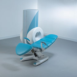
From ONI Medical Systems, Inc.;
MSK-Extreme™ MRI system is a dedicated high field extremity imaging device, designed to provide orthopedic surgeons and other physicians with detailed diagnostic images of the foot, ankle, knee, hand, wrist and elbow, all with the clinical confidence and advantages derived from high field, whole body MRI units. The light weight (less than 650 kg) of the OrthOne System performs rapid patient studies, is easy to operate, has a patient friendly open environment and can be installed in a practice office or hospital, all at a cost similar to a low field extremity machine.
New features include a more powerful operating system that offers increased scan speed as well as a 160-mm knee coil with higher signal to noise ratio, and the option of a CD burner.
Device Information and Specification 16 cm knee, 18 cm lower extremity;; 12.3 cm upper extremity, additional high resolution v-SPEC Coils: 80 mm, 100 mm, or 145 mm. SE, FSE, GE2D, GE3D, Inversion recovery (IR), Driven Equilibrium, Fat Saturation (FS), STIR, MT, PD, Flow Compensation (FC), RF spoiling, MTE, No Phase Wrap (NPW) IMAGING MODES Scout, single, multislice, volume 2D less than 200 msec/image X/Y: 64-512; 2 pixel steps 4,096 grey lvls; 256 lvls in 3D POWER REQUIREMENTS 115VAC, 1phase, 20A; 208VAC, 3 phase, 30A COOLING SYSTEM TYPE LHe with 2 stage cold head 1.25m radial x 1.8m axial | |  | | | |  Further Reading: Further Reading: | Basics:
|
|
| |
|  | |  |  |
|  | |
|  | 1 - 5 (of 8) nextResult Pages :  [1] [1]  [2] [2] |
| |
|
| |
 | Look
Ups |
| |