Magnetic Resonance - Technology Information Portal Welcome to MRI Technology
Info
  Sheets

Out-
      side


 'Rephasing Gradient' 
SEARCH FOR    
 
  2 3 5 A B C D E F G H I J K L M N O P Q R S T U V W X Y Z
Result : Searchterm 'Rephasing Gradient' found in 1 term [] and 0 definition [], (+ 9 Boolean[] results
1 - 5 (of 10)     next
Result Pages : [1]  [2]
MRI Resources 
Safety Training - Spectroscopy pool - Societies - Spine MRI - Non-English - Homepages
 
Rephasing Gradient
 
Magnetic field gradient pulse applied to reverse the spatial variation of phase of transverse magnetization caused by a dephasing gradient. For example, in selective excitation, it is a magnetic field gradient applied for a brief period after a selective excitation pulse, in the opposite direction to the gradient used for the selective excitation. The result of the gradient reversal is a rephasing of the spins (which will have gotten out of phase with each other along the direction of the selection gradient), forming a gradient echo and improving the sensitivity of imaging after the selective excitation process.
spacer
 
• Share the entry 'Rephasing Gradient':  Facebook  Twitter  LinkedIn  
MRI Resources 
Distributors - Health - Collections - Sequences - Process Analysis - Online Books
 
Gradient Motion Rephasing
 
(GMR) The application of strategic gradient pulses can compensate the objectionable spin phase effects of flow motion. That means the reducing of flow effects, e.g. gradient moment nulling of the first order of flow. The simplest velocity-compensated pulse sequence is the symmetrical second echo of a spin echo pulse sequence.
Gradient field changes can be configured in such a way that during an echo the magnetization signal vectors for all pixels have zero phase angle independent of velocities, accelerations etc. of the measured tissue. E.g. the adjustment to zero at the time TE of the net moments of the amplitude of the waveform of the magnetic field gradients with time. The zeroth moment is the area under the curve, the first moment is the 'center of gravity' etc. The aim is to minimize the phase shifts acquired by the transverse magnetization of excited nuclei moving along the gradients (including the effect of refocusing RF pulses), particularly for the reduction of image artifacts due to motion.
Also called Flow Compensation (FC), Motion Artifact Suppression Technique (MAST), Flow motion compression (STILL), Gradient Rephasing (GR), Shimadzu Motion Artifact Reduction Technique (SMART).
spacer

• View the DATABASE results for 'Gradient Motion Rephasing' (2).Open this link in a new window

 
Further Reading:
  Basics:
Motion Compensation in MR Imaging
   by ccn.ucla.edu    
MRI Resources 
Pathology - Movies - Equipment - Crystallography - Corporations - Spectroscopy
 
Fast Spin EchoForum -
related threadsInfoSheet: - Sequences - 
Intro, 
Overview, 
Types of, 
etc.
 
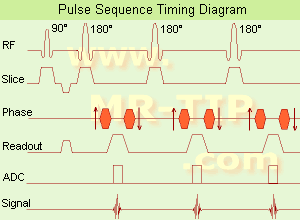
Fast Spin Echo Diagram (FSE) In the pulse sequence timing diagram, a fast spin echo sequence with an echo train length of 3 is illustrated. This sequence is characterized by a series of rapidly applied 180° rephasing pulses and multiple echoes, changing the phase encoding gradient for each echo.
The echo time TE may vary from echo to echo in the echo train. The echoes in the center of the K-space (in the case of linear k-space acquisition) mainly produce the type of image contrast, whereas the periphery of K-space determines the spatial resolution. For example, in the middle of K-space the late echoes of T2 weighted images are encoded. T1 or PD contrast is produced from the early echoes.
The benefit of this technique is that the scan duration with, e.g. a turbo spin echo turbo factor / echo train length of 9, is one ninth of the time. In T1 weighted and proton density weighted sequences, there is a limit to how large the ETL can be (e.g. a usual ETL for T1 weighted images is between 3 and 7). The use of large echo train lengths with short TE results in blurring and loss of contrast. For this reason, T2 weighted imaging profits most from this technique.
In T2 weighted FSE images, both water and fat are hyperintense. This is because the succession of 180° RF pulses reduces the spin spin interactions in fat and increases its T2 decay time. Fast spin echo (FSE) sequences have replaced conventional T2 weighted spin echo sequences for most clinical applications. Fast spin echo allows reduced acquisition times and enables T2 weighted breath hold imaging, e.g. for applications in the upper abdomen.
In case of the acquisition of 2 echoes this type of a sequence is named double fast spin echo / dual echo sequence, the first echo is usually density and the second echo is T2 weighted image. Fast spin echo images are more T2 weighted, which makes it difficult to obtain true proton density weighted images. For dual echo imaging with density weighting, the TR should be kept between 2000 - 2400 msec with a short ETL (e.g., 4).
Other terms for this technique are:
Turbo Spin Echo
Rapid Imaging Spin Echo,
Rapid Spin Echo,
Rapid Acquisition Spin Echo,
Rapid Acquisition with Refocused Echoes
 
Images, Movies, Sliders:
 Lumbar Spine T2 FSE Sagittal  Open this link in a new window
    

Courtesy of  Robert R. Edelman
 MRI - Anatomic Imaging of the Foot  Open this link in a new window
    
SlidersSliders Overview

 Lumbar Spine T2 FSE Axial  Open this link in a new window
    

Courtesy of  Robert R. Edelman
 
spacer

• View the DATABASE results for 'Fast Spin Echo' (31).Open this link in a new window

 
Further Reading:
  Basics:
MYELIN-SELECTIVE MRI: PULSE SEQUENCE DESIGN AND OPTIMIZATION
   by www.imaging.robarts.ca    
Advances in Magnetic Resonance Neuroimaging
Friday, 27 February 2009   by www.ncbi.nlm.nih.gov    
  News & More:
New MR sequence helps radiologists more accurately evaluate abnormalities of the uterus and ovaries
Thursday, 23 April 2009   by www.eurekalert.org    
Spin echoes, CPMG and T2 relaxation - Introductory NMR & MRI from Magritek
2013   by www.azom.com    
MRI Resources 
MRI Technician and Technologist Jobs - Software - Safety Training - Bioinformatics - Guidance - Health
 
Motion Artifact Suppression TechniqueMRI Resource Directory:
 - Artifacts -
 
spacer

• View the DATABASE results for 'Motion Artifact Suppression Technique' (3).Open this link in a new window

 
Further Reading:
  News & More:
KinetiCor Wins FDA 510(k) Clearance for Motion Correction System for Siemens MAGNETOM Skyra 3T Scanner
Wednesday, 19 February 2020   by finance.yahoo.com    
Chest MRI Using Multivane-XD, a Novel T2-Weighted Free Breathing MR Sequence
Thursday, 11 July 2019   by www.sciencedirect.co    
Prospective motion correction using coil-mounted cameras: Cross-calibration considerations
Saturday, 7 April 2018   by www.ncbi.nlm.nih.gov    
T2-Weighted Liver MRI Using the MultiVane Technique at 3T: Comparison with Conventional T2-Weighted MRI
Friday, 16 October 2015   by www.ncbi.nlm.nih.gov    
MRI Resources 
Liver Imaging - Services and Supplies - MRI Reimbursement - Case Studies - Musculoskeletal and Joint MRI - MRI Accidents
 
Spin EchoForum -
related threads
 
(SE) The Reappearance of the MR signal after the FID has apparently died away, as a result of the effective reversal (rephasing) of the dephasing spins by techniques such as specific RF pulse sequences or pairs of field gradient pulses, applied in time shorter than or on the order of T2. Proper selection of the TE time of the pulse sequence can help to control the amount of T1 or T2 contrast present in the image. Pulse sequences of the spin echo type, usually employs a 90° pulse, followed by one or more 180° pulses to eliminate field inhomogeneity and chemical shift effects at the echo. Caused by this 180° refocusing pulse, spin echo or fast spin echo (FSE, TSE) sequences are more robust against e.g. susceptibility artifacts than sequences of the gradient echo type.
spacer

• View the DATABASE results for 'Spin Echo' (96).Open this link in a new window


• View the NEWS results for 'Spin Echo' (1).Open this link in a new window.
 
Further Reading:
  Basics:
Spin echoes, CPMG and T2 relaxation - Introductory NMR & MRI from Magritek
2013   by www.azom.com    
  News & More:
EVALUATION OF HUMAN STROKE BY MR IMAGING
2000
MRI Resources 
Portals - Online Books - Liver Imaging - Mobile MRI Rental - Directories - Safety Training
 
     1 - 5 (of 10)     next
Result Pages : [1]  [2]
 Random Page
 
Share This Page
FacebookTwitterLinkedIn

MR-TIP    
Community   
User
Pass
Forgot your UserID/Password ?    


Personalized protocols (age, gender, body habitus, etc.) lead to :
more automated planning 
improved patient comfort 
better diagnostics 
optimized image quality 
nothing 

Look
      Ups


MR-TIP.com uses cookies! By browsing MR-TIP.com, you agree to our use of cookies.

Magnetic Resonance - Technology Information Portal
Member of SoftWays' Medical Imaging Group - MR-TIP • Radiology-TIP • Medical-Ultrasound-Imaging • 
Copyright © 2003 - 2024 SoftWays. All rights reserved. [ 21 November 2024]
Terms of Use | Privacy Policy | Advertising
 [last update: 2024-02-26 03:41:00]