 | Info
Sheets |
| | | | | | | | | | | | | | | | | | | | | | | | |
 | Out-
side |
| | | | |
|
| | | | |
Result : Searchterm 'SAR' found in 3 terms [ ] and 60 definitions [ ] and 60 definitions [ ] ]
| previous 16 - 20 (of 63) nextResult Pages :  [1] [1]  [2 3 4 5 6 7 8 9 10 11 12 13] [2 3 4 5 6 7 8 9 10 11 12 13] |  | |  | Searchterm 'SAR' was also found in the following services: | | | | |
|  |  |
| |
|

From Hitachi Medical Systems America, Inc.;
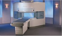
the AIRIS made its debut in 1995. Hitachi followed up with the AIRIS II system, which has proven equally successfully. 'All told, Hitachi has installed more than 1,000 MRI systems in the U.S., holding more than 17 percent of the total U.S. MRI installed base, and more than half of the installed base of open MR systems,' says Antonio Garcia, Frost and Sullivan industry research analyst.
Now Altaire employs a blend of innovative Hitachi features called VOSI™ technology, optimizing each sub-system's performance in concert with the
other sub-systems, to give the seamless mix of high-field performance
and the patient comfort, especially for claustrophobic patients, of open MR systems.
Device Information and Specification
CLINICAL APPLICATION
Whole body
DualQuad T/R Body Coil, MA Head, MA C-Spine, MA Shoulder, MA Wrist, MA CTL Spine, MA Knee, MA TMJ, MA Flex Body (3 sizes), Neck, small and large Extremity, PVA (WIP), Breast (WIP), Neurovascular (WIP), Cardiac (WIP) and MA Foot//Ankle (WIP)
SE, GE, GR, IR, FIR, STIR, ss-FSE, FSE, DE-FSE/FIR, FLAIR, ss/ms-EPI, ss/ms EPI- DWI, SSP, MTC, SE/GE-EPI, MRCP, SARGE, RSSG, TRSG, BASG, Angiography: CE, PC, 2D/3D TOF
IMAGING MODES
Single, multislice, volume study
TR
SE: 30 - 10,000msec GE: 3.6 - 10,000msec IR: 50 - 16,700msec FSE: 200 - 16,7000msec
TE
SE : 8 - 250msec IR: 5.2 -7,680msec GE: 1.8 - 2,000 msec FSE: 5.2 - 7,680
0.05 sec/image (256 x 256)
2D: 2 - 100 mm; 3D: 0.5 - 5 mm
Level Range: -2,000 to +4,000
COOLING SYSTEM TYPE
Water-cooled
3.1 m lateral, 3.6 m vertical
| |  | | | | | | | | |  Further Reading: Further Reading: | News & More:
|
|
| |
|  | |  |  |  |
| |
|
| | | |  | |
• View the DATABASE results for 'Angiography' (120).
| | |
• View the NEWS results for 'Angiography' (15).
| | | | |  Further Reading: Further Reading: | News & More:
|
|
| |
|  | |  |  |  |
| |
|
This family of sequences uses a balanced gradient waveform. This waveform will act on any stationary spin on resonance between 2 consecutive RF pulses and return it to the same phase it had before the gradients were applied.
A balanced sequence starts out with a RF pulse of 90° or less and the spins in the steady state. Prior to the next TR in the slice encoding, the phase encoding and the frequency encoding direction, gradients are balanced so their net value is zero. Now the spins are prepared to accept the next RF pulse, and their corresponding signal can become part of the new transverse magnetization. If the balanced gradients maintain the longitudinal and transverse magnetization, the result is that both T1 and T2 contrast
are represented in the image.
This pulse sequence produces images with increased signal from fluid (like T2 weighted sequences), along with retaining T1 weighted tissue contrast. Balanced sequences are particularly useful in cardiac MRI. Because this form of sequence is extremely dependent on field homogeneity, it is essential to run a shimming prior the acquisition.
Usually the gray and white matter contrast is poor, making this type of sequence unsuited for brain MRI. Modifications like ramping up and down the flip angles can increase signal to noise ratio and contrast of brain tissues (suggested under the name COSMIC - Coherent Oscillatory State acquisition for the Manipulation of Image Contrast).
These sequences include e.g. Balanced Fast Field Echo (bFFE), Balanced Turbo Field Echo ( bTFE), Fast Imaging with Steady Precession ( TrueFISP, sometimes short TRUFI), Completely Balanced Steady State (CBASS) and Balanced SARGE (BASG). | | | |  | |
• View the DATABASE results for 'Balanced Sequence' (5).
| | | | |  Further Reading: Further Reading: | News & More:
|
|
| |
|  |  | Searchterm 'SAR' was also found in the following services: | | | | |
|  |  |
| |
|
Eisai Co., Ltd. (Tokyo, Japan, President: Haruo Naito) and Bracco-Eisai Co., Ltd. (Tokyo, Japan, President: Toshio Matsumoto) decided to withdraw the new drug application (NDA) of E7155 (gadobenate meglumine), since new indications are emerging for the product and additional clinical data was necessary to complete the targeting product competitive risk/benefit profile. The product is currently marketed in other countries, and both companies are discussing a future development plan in Japan.
MRI Contrast Agents:
Contact Information
MAIL
Bracco Eisai Co., Ltd.
2-6, Kohinata 4-chome, Bunkyo-ku, Tokyo, 112-0006
| |  | |
• View the DATABASE results for 'Bracco-Eisai Co., Ltd.' (2).
| | | | |
|  | |  |  |  |
| |
|
A pacemaker is a device for internal or external battery-operated cardiac pacing to overcome cardiac arrhythmias or heart block. All implanted electronic devices are susceptible to the electromagnetic fields used in magnetic resonance imaging. Therefore, the main magnetic field, the gradient field, and the radio frequency (RF) field are potential hazards for cardiac pacemaker patients.
The pacemaker's susceptibility to static field and its critical role in life support have warranted special consideration. The static magnetic field applies force to magnetic materials. This force and torque effects rise linearly with the field strength of the MRI machines. Both, RF fields and pulsed gradients can induce voltages in circuits or on the pacing lead, which will heat up the tissue around e.g. the lead tip, with a potential risk of thermal injury.
Regulations for pacemakers provide that they have to switch to the magnet mode in static magnetic fields above 1.0 mT. In MR imaging, the gradient and RF fields may mimic signals from the heart with inhibition or fast pacing of the heart. In the magnet mode, most of the current pacemakers will pace with a fix pulse rate because they do not accept the heartsignals. However, the state of an implanted pacemaker will be unpredictable inside a strong magnetic field. Transcutaneous controller adjustment of pacing rate is a feature of many units. Some achieve this control using switches activated by the external application of a magnet to open/close the switch. Others use rotation of an external magnet to turn internal controls. The fringe field around the MRI magnet can activate such switches or controls. Such activations are a safety risk.
Areas with fields higher than 0.5 mT ( 5 Gauss Limit) commonly have restricted access and/or are posted as a safety risk to persons with pacemakers.

A Cardiac pacemaker is because the risks, under normal circumstances an absolute contraindication for MRI procedures.
Nevertheless, with special precaution the risks can be lowered. Reprogramming the pacemaker to an asynchronous mode with fix pacing rate or turning off will reduce the risk of fast pacing or inhibition. Reducing the SAR value reduces the potential MRI risks of heating. For MRI scans of the head and the lower extremities, tissue heating also seems to be a smaller problem. If a transmit receive coil is used to scan the head or the feet, the cardiac pacemaker is outside the sending coil and possible heating is very limited. | |  | |
• View the DATABASE results for 'Cardiac Pacemaker' (6).
| | | | |  Further Reading: Further Reading: | | Basics:
|
|
News & More:
| |
| |
|  | |  |  |
|  | |
|  | | |
|
| |
 | Look
Ups |
| |