Magnetic Resonance - Technology Information Portal Welcome to MRI Technology
Info
  Sheets

Out-
      side


 'SPIR' 
SEARCH FOR    
 
  2 3 5 A B C D E F G H I J K L M N O P Q R S T U V W X Y Z
Result : Searchterm 'SPIR' found in 8 terms [] and 81 definitions []
previous     46 - 50 (of 89)     next
Result Pages : [1 2]  [3 4 5 6 7 8 9 10 11 12 13 14 15 16 17 18]
Searchterm 'SPIR' was also found in the following services: 
spacer
News  (17)  Resources  (5)  Forum  (8)  
 
Excelart XG™ with PianissimoInfoSheet: - Devices -
Intro, 
Types of Magnets, 
Overview, 
etc.MRI Resource Directory:
 - Devices -
 
http://www.medical.toshiba.com/clinical/radiology/15texcelart.htm From Toshiba America Medical Systems Inc.;
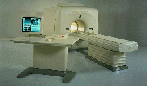
the EXCELART is a superconducting whole body MRI system with a short wide-bore magnet, operating at 1.5 T. It features powerful high-speed gradients with a revolutionary gradient acoustic noise reduction system: Pianissimo. The dramatic reduction of gradient acoustic noise by Pianissimo greatly enhances patient comfort during exams. The standard array platform and a wide range of array coils ensure excellent images. A powerful 64-bit RISC-based computer system and newly developed array processor realize high productivity.
Device Information and Specification
CLINICAL APPLICATION
Whole body
CONFIGURATION
Cylindrical Wide Short Bore
Optional (WIP)
SYNCHRONIZATION
ECG/peripheral, respiratory gating
PULSE SEQUENCES
SE, FE, IR, FastSE, FastIR, FastFLAIR, Fast STIR, FastFE, FASE, Hybrid EPI, Multi Shot EPI; Angiography: 2D(gate/non-gate)/3D TOF, SORS-STC
IMAGING MODES
Single, multislice, volume study
TR
2.6-30000 msec
TE
8 msec min. SE; 0.9 msec min. FE
SINGLE/MULTI SLICE
less than 0.011 (256x256)
FOV
2 cm
1.0 min. 2-DFT: 0.2 min. 3-DFT
Up to 1024
MEASURING MATRIX
32-1024, phase;; 64-1024, freq.
PIXEL INTENSITY
256 gray levels
BORE DIAMETER
or W x H
65.5 cm, patient aperture
MAGNET WEIGHT
4050 kg (bare magnet incl. L-He)
H*W*D
235 x 219 x 199 cm
POWER REQUIREMENTS
380/400/415/440/480 V
COOLING SYSTEM TYPE
Closed-loop water-cooled
CRYOGEN USE
Liquid helium: approx. less than 0.05 L/hr
STRENGTH
25 mT/m
5-GAUSS FRINGE FIELD
2.5 m / 4.0 m
Passive, active, auto-active
spacer
 
Further Reading:
  News & More:
Angio, cardiac imaging top list for MR and CT
1999   by www.diagnosticimaging.com    
MRI Resources 
MRI Training Courses - Stimulator pool - Mobile MRI Rental - Databases - Stent - Shielding
 
FLEXART™InfoSheet: - Devices -
Intro, 
Types of Magnets, 
Overview, 
etc.MRI Resource Directory:
 - Devices -
 
From Toshiba America Medical Systems Inc.; www3.toshiba.co.jp/medical/products/mr/flex/index.htm FLEXART™ series is a 0.5 T superconducting MRI system that has been designed to meet the expanding role of MRI in today's clinical environment. The system utilizes innovative technologies such as digital RF, high speed actively shielded gradients and optimized RF coils which support a wide range of MRI developments.
Device Information and Specification
CLINICAL APPLICATION
Whole body
CONFIGURATION
Compact
Quadrature, solenoid and multi-channel configurations
SYNCHRONIZATION
ECG/peripheral optional, respiratory gating
PULSE SEQUENCES
SE, FE, IR, FastSE, FastIR, FastFLAIR, Fast STIR, FastFE, FASE, Hybrid EPI, Multi Shot EPI; Angiography: 2D(gate/non-gate)/3D TOF, SORS-STC
IMAGING MODES
Single, multislice, volume study
POWER REQUIREMENTS
380/400/415/440/480 V
COOLING SYSTEM TYPE
Closed-loop water-cooled
STRENGTH
25 mT/m
Passive, active
spacer

• View the DATABASE results for 'FLEXART™' (2).Open this link in a new window

MRI Resources 
Cardiovascular Imaging - Health - Stimulator pool - MRI Accidents - Brain MRI - Breast Implant
 
FORTE 3.0T™InfoSheet: - Devices -
Intro, 
Types of Magnets, 
Overview, 
etc.MRI Resource Directory:
 - Devices -
 
www.isoltech.co.kr/english/product/30t.htm From ISOL Technology
'Ultra high field MR system, it's right close to you. FORTE 3.0T is the new standard for the future ultra high field MR system. If you are pushing the limits of your existing clinical MR scanner, the FORTE will surely take you to the next level of diagnostic imaging. FORTE is the core leader of the medical technology in the 21st century. Proving effects of fMRI that cannot be measured with MRI less than 2.0T.'
Device Information and Specification
CLINICAL APPLICATION
Whole body
CONFIGURATION
Short bore compact
SYNCHRONIZATION
ECG/peripheral: Optional/yes, external trigger, respiratory gating
PULSE SEQUENCES
Spin echo, Gradient echo, Fast spin echo, Inversion recovery, 2D/3D Fast gradient echo sequences FLAIR/STIR, 2D/3D TOF
IMAGING MODES
2D/3D, T1, T2 and Diffusion//Perfusion imaging, MR Angiography package, Advanced EPI package, Multi-nuclei MR Spectroscopy package
FOV
40 cm
128 x 128, 256 x 256, 512 x 512, 1024 x 1024
BORE DIAMETER
or W x H
61 cm without body coil
MAGNET WEIGHT
12000 kg
H*W*D
260 x 220 x 235 cm
COOLING SYSTEM TYPE
Water-cooled coil and air-cooled amplifier
CRYOGEN USE
0.15 L/hr helium
STRENGTH
38 mT/m
5-GAUSS FRINGE FIELD
3.3 m / 5.2 m
Passive and active
spacer

• View the DATABASE results for 'FORTE 3.0T™' (2).Open this link in a new window

Searchterm 'SPIR' was also found in the following services: 
spacer
News  (17)  Resources  (5)  Forum  (8)  
 
Fast Relaxation Fast Spin EchoInfoSheet: - Sequences - 
Intro, 
Overview, 
Types of, 
etc.
 
(FRFSE, FR-FSE) The fast relaxation fast spin echo sequence provides high signal intensity of fluids even with short repetition times, and can be used with parallel imaging techniques for short breath hold imaging or respiratory gating for free-breathing, high isotropic resolution MR imaging. After signal decay at the end of the echo train, a negative 90° pulse align spins with long T2 from the transverse plane to the longitudinal plane, leading to a much faster recovery of tissues with long T2 time to the equilibrium and thus better contrast between tissues with long and short T2.
Fast relaxation FSE has advantages also for volumetric imaging as the TR can be substantially reduced and thus the scan time. The sequence can be post processed with maximum intensity projection, surface or volume rendering algorithms to visualize anatomical details in brain or spine MRI. Cerebro spinal fluid pulsation artifacts, often problematic in the cervical or thoracic spine may be reduced by radial sampling, in particular when combined with acquisitions of the PROPELLER type.

See also Fast spin echo, Driven Equilibrium.
 
Images, Movies, Sliders:
 Shoulder Sagittal T2 FatSat FRFSE  Open this link in a new window
    

Courtesy of  Robert R. Edelman
 Shoulder Axial T2 FatSat FRFSE  Open this link in a new window
 Shoulder Coronal T2 FatSat FRFSE  Open this link in a new window
    

Courtesy of  Robert R. Edelman
 
spacer
MRI Resources 
Supplies - DICOM - Calculation - Education pool - Nerve Stimulator - Implant and Prosthesis pool
 
FlowForum -
related threads
 
Flow phenomena are intrinsic processes in the human body. Organs like the heart, the brain or the kidneys need large amounts of blood and the blood flow varies depending on their degree of activity. Magnetic resonance imaging has a high sensitivity to flow and offers accurate, reproducible, and noninvasive methods for the quantification of flow. MRI flow measurements yield information of blood supply of of various vessels and tissues as well as cerebro spinal fluid movement.
Flow can be measured and visualized with different pulse sequences (e.g. phase contrast sequence, cine sequence, time of flight angiography) or contrast enhanced MRI methods (e.g. perfusion imaging, arterial spin labeling).
The blood volume per time (flow) is measured in: cm3/s or ml/min. The blood flow-velocity decreases gradually dependent on the vessel diameter, from approximately 50 cm per second in arteries with a diameter of around 6 mm like the carotids, to 0.3 cm per second in the small arterioles.

Different flow types in human body:
Behaves like stationary tissue, the signal intensity depends on T1, T2 and PD = Stagnant flow
Flow with consistent velocities across a vessel = Laminar flow
Laminar flow passes through a stricture or stenosis (in the center fast flow, near the walls the flow spirals) = Vortex flow
Flow at different velocities that fluctuates = Turbulent flow

See also Flow Effects, Flow Artifact, Flow Quantification, Flow Related Enhancement, Flow Encoding, Flow Void, Cerebro Spinal Fluid Pulsation Artifact, Cardiovascular Imaging and Cardiac MRI.
 
Images, Movies, Sliders:
 MVP Parasternal  Open this link in a new window
    

Courtesy of  Robert R. Edelman
 TOF-MRA Circle of Willis Inverted MIP  Open this link in a new window
    

 Circle of Willis, Time of Flight, MIP  Open this link in a new window
    
SlidersSliders Overview

 
spacer

• View the DATABASE results for 'Flow' (113).Open this link in a new window


• View the NEWS results for 'Flow' (7).Open this link in a new window.
 
Further Reading:
  News & More:
The super-fast MRI scan that could revolutionise heart failure diagnosis
Wednesday, 21 September 2022   by www.eurekalert.org    
MRI Resources 
Fluorescence - Directories - Mass Spectrometry - NMR - Claustrophobia - Chemistry
 
previous      46 - 50 (of 89)     next
Result Pages : [1 2]  [3 4 5 6 7 8 9 10 11 12 13 14 15 16 17 18]
 Random Page
 
Share This Page
FacebookTwitterLinkedIn

MR-TIP    
Community   
User
Pass
Forgot your UserID/Password ?    


Personalized protocols (age, gender, body habitus, etc.) lead to :
more automated planning 
improved patient comfort 
better diagnostics 
optimized image quality 
nothing 

Look
      Ups


MR-TIP.com uses cookies! By browsing MR-TIP.com, you agree to our use of cookies.

Magnetic Resonance - Technology Information Portal
Member of SoftWays' Medical Imaging Group - MR-TIP • Radiology-TIP • Medical-Ultrasound-Imaging • 
Copyright © 2003 - 2024 SoftWays. All rights reserved. [ 23 November 2024]
Terms of Use | Privacy Policy | Advertising
 [last update: 2024-02-26 03:41:00]