 | Info
Sheets |
| | | | | | | | | | | | | | | | | | | | | | | | |
 | Out-
side |
| | | | |
|
| | | | | |  | Searchterm 'SPIR' was also found in the following services: | | | | |
|  |  |
| |
|
Quick Overview
HELP
cardiac and re spiratory synchronization
Movement of the heart causes blurring and ghosting in the images. The artifacts appear in the phase encoding direction, independent of the direction of the motion.

Image Guidance
These artifacts can be reduced by using cardiac synchronization: triggering, gating or retrospective triggering. Maximum reduction can be achieved by using triggering in combination with flow compensation, re spiratory triggering or breath hold and regional saturation techniques.
See also Motion Artifact. | | | |  | | | |  Further Reading: Further Reading: | | Basics:
|
|
News & More:
| |
| |
|  | |  |  |  |
| |
|
Fat suppression is the process of utilizing specific MRI parameters to remove the deleterious effects of fat from the resulting images , e.g. with STIR, FAT SAT sequences, water selective (PROSET WATS - water only selection, also FATS - fat only selection possible) excitation techniques, or pulse sequences based on the Dixon method.
Spin magnetization can be modulated by using special RF pulses. CHESS or its variations like SPIR, SPAIR ( Spectral Selection Attenuated Inversion Recovery) and FAT SAT use frequency selective excitation pulses, which produce fat saturation.
Fat suppression techniques are nearly used in all body parts and belong to every standard MRI protocol of joints like knee, shoulder, hips, etc.

Image Guidance
Imaging of, e.g. the foot can induce bad fat suppression with SPIR/FAT SAT due to the asymmetric volume of this body part. The volume of the foot alters the magnetic field to a different degree than the smaller volume of the lower leg affecting the protons there. There is only a small band of tissue where the fat protons are precessing at the frequency expected, resulting in frequency selective fat saturation working only in that area. This can be corrected by volume shimming or creating a more symmetrical volume being imaged with water bags.
Even with their longer scan time and motion sensitivity, STIR (short T1/tau inversion recovery) sequences are often the better choice to suppress fat. STIR images are also preferred because of the decreased sensitivity to field inhomogeneities, permitting larger fields of views when compared to fat suppressed images and the ability to image away from the isocenter. See also Knee MRI.
Sequences based on Dixon turbo spin echo ( fast spin echo) can deliver a significant better fat suppression than conventional TSE/FSE imaging.
| | | |  | |
• View the DATABASE results for 'Fat Suppression' (28).
| | | | |  Further Reading: Further Reading: | | Basics:
|
|
News & More:
| |
| |
|  | |  |  |  |
| |
|
Device Information and Specification
CLINICAL APPLICATION
Whole body
CONFIGURATION
Short bore compact
Standard: head, body, C1, C3; Optional: Small joint, flex-E, flex-R, endocavitary (L and S), dual TMJ, knee, neck, T/L spine, breast; Optional phased array: Spine, pediatric, 3rd party connector, Flex-S-M-L, flex body, flex cardiac, neuro-vascular, head
SE, Modified-SE ( TSE), DAVE, STIR, FLAIR, SPIR, MTC, Dynamic, Keyhole, CLEAR, Q Flow, Balanced FFE, Multi Chunk 3D, Multi Stack 3D, FFE-EPI, SE-EPI, IR-EPI, GRASE, Diffusion Imaging, Perfusion Imaging;; Angiography: Inflow MRA, TONE, PCA, CE MRA
RapidView Recon. greater than 500 @ 256 Matrix
128 x 128, 256 x 256,512 x 512,1024 x 1024
Variable in 1% increments
Lum.: 120 cd/m2; contrast: 150:1
Variable (op. param. depend.)
POWER REQUIREMENTS
380/400 V
| |  | |
• View the DATABASE results for 'Intera 0.5T™' (2).
| | | | |
|  |  | Searchterm 'SPIR' was also found in the following services: | | | | |
|  |  |
| |
|
Device Information and Specification
CLINICAL APPLICATION
Whole body
CONFIGURATION
Short bore compact
Standard: head, body, C1, C3; Optional: Small joint, flex-E, flex-R, endocavitary (L and S), dual TMJ, knee, neck, T/L spine, breast; Optional phased array: Spine, pediatric, 3rd party connector, Optional SENSE Coils: Flex-S-M-L, Flex Body, Flex Cardiac
SE, Modified-SE, IR (T1, T2, PD), STIR, FLAIR, SPIR, FFE, T1-FFE, T2-FFE, Balanced FFE, TFE, Balanced TFE, Dynamic, Keyhole, 3D, Multi Chunk 3D, Multi Stack 3D, K Space Shutter, MTC, TSE, Dual IR, DRIVE, EPI, Cine, 2DMSS, DAVE, Mixed Mode; Angiography: Inflow MRA, TONE, PCA, CE MRA
TR
Min. 2.9 (Omni) msec, 1.6 (Power) msec
TE
Min. 1.0 (Omni) msec, 0.7 (Power) msec
RapidView Recon. greater than 500 @ 256 Matrix
0.1 mm(Omni), 0.05 mm (Power)
128 x 128, 256 x 256,512 x 512,1024 x 1024 (64 for Bold img)
Variable in 1% increments
Lum.: 120 cd/m2; contrast: 150:1
Variable (op. param. depend.)
POWER REQUIREMENTS
380/400 V
STRENGTH
23 mT/m (Omni), 30 (Power) mT/m
| |  | |
• View the DATABASE results for 'Intera 1.0T™' (2).
| | | | |
|  | |  |  |  |
| |
|

From Philips Medical Systems;
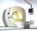
the Intera-family offers with this member a wide range of possibilities, efficiency and a ergonomic and intuitive serving-platform. Also available as Intera CV for cardiac and Intera I/T for interventional MR procedures.
The scanners are also equipped with SENSE technology, which is essential for high-quality contrast enhanced magnetic resonance angiography, interactive cardiac MR and diffusion tensor imaging ( DTI) fiber tracking.
The increased accuracy and clarity of MR scans obtained with this technology allow for faster and more accurate diagnosis of potential problems like patient friendliness and expands the breadth of applications including cardiology, oncology and interventional MR.
Device Information and Specification
CLINICAL APPLICATION
Whole body
CONFIGURATION
Short bore compact
Standard: head, body, C1, C3; Optional: Small joint, flex-E, flex-R, endocavitary (L and S), dual TMJ, knee, neck, T/L spine, breast; Optional phased array: Spine, pediatric, 3rd party connector; Optional SENSE coils: Flex-S-M-L, flex body, flex cardiac
SE, Modified-SE ( TSE), IR (T1, T2, PD), STIR, FLAIR, SPIR, FFE, T1-FFE, T2-FFE, Balanced FFE, TFE, Balanced TFE, Dynamic, Keyhole, 3D, Multi Chunk 3D, Multi Stack 3D, K Space Shutter, MTC, TSE, Dual IR, DRIVE, EPI, Cine, 2DMSS, DAVE, Mixed Mode; Angiography: PCA, MCA, Inflow MRA, CE
TR
2.9 (Omni), 1.6 (Power), 1.6 (Master/Expl) msec
TE
1.0 (Omni), 0.7 (Power), 0.5 (Master/Expl) msec
RapidView Recon. greater than 500 @ 256 Matrix
0.1 mm(Omni), 0.05 mm (Pwr/Mstr/Expl)
128 x 128, 256 x 256,512 x 512,1024 x 1024 (64 for BOLD img.)
Variable in 1% increments
Lum.: 120 cd/m2; contrast: 150:1
Variable (op. param. depend.)
POWER REQUIREMENTS
380/400 V
| |  | |
• View the DATABASE results for 'Intera 1.5T™' (2).
| | | | |
|  | |  |  |
|  | | |
|
| |
 | Look
Ups |
| |