Magnetic Resonance - Technology Information Portal Welcome to MRI Technology
Info
  Sheets

Out-
      side


 'SPIR' 
SEARCH FOR    
 
  2 3 5 A B C D E F G H I J K L M N O P Q R S T U V W X Y Z
Result : Searchterm 'SPIR' found in 8 terms [] and 81 definitions []
previous     36 - 40 (of 89)     next
Result Pages : [1 2]  [3 4 5 6 7 8 9 10 11 12 13 14 15 16 17 18]
Searchterm 'SPIR' was also found in the following services: 
spacer
News  (17)  Resources  (5)  Forum  (8)  
 
Altaireâ„¢InfoSheet: - Devices -
Intro, 
Types of Magnets, 
Overview, 
etc.MRI Resource Directory:
 - Devices -
 
www.hitachimed.com/products/altaire.asp From Hitachi Medical Systems America, Inc.;
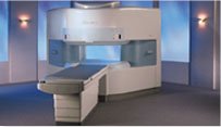
the AIRIS made its debut in 1995. Hitachi followed up with the AIRIS II system, which has proven equally successfully. 'All told, Hitachi has installed more than 1,000 MRI systems in the U.S., holding more than 17 percent of the total U.S. MRI installed base, and more than half of the installed base of open MR systems,' says Antonio Garcia, Frost and Sullivan industry research analyst. Now Altaire employs a blend of innovative Hitachi features called VOSIâ„¢ technology, optimizing each sub-system's performance in concert with the other sub-systems, to give the seamless mix of high-field performance and the patient comfort, especially for claustrophobic patients, of open MR systems.

Device Information and Specification
CLINICAL APPLICATION
Whole body
CONFIGURATION
Vertical Field, Open MRI
DualQuad T/R Body Coil, MA Head, MA C-Spine, MA Shoulder, MA Wrist, MA CTL Spine, MA Knee, MA TMJ, MA Flex Body (3 sizes), Neck, small and large Extremity, PVA (WIP), Breast (WIP), Neurovascular (WIP), Cardiac (WIP) and MA Foot//Ankle (WIP)
SYNCHRONIZATION
Cardiac gating, ECG/peripheral, respiratory gating (2 modes)
PULSE SEQUENCES
SE, GE, GR, IR, FIR, STIR, ss-FSE, FSE, DE-FSE/FIR, FLAIR, ss/ms-EPI, ss/ms EPI- DWI, SSP, MTC, SE/GE-EPI, MRCP, SARGE, RSSG, TRSG, BASG, Angiography: CE, PC, 2D/3D TOF
IMAGING MODES
Single, multislice, volume study
TR
SE: 30 - 10,000msec GE: 3.6 - 10,000msec IR: 50 - 16,700msec FSE: 200 - 16,7000msec
TE
SE : 8 - 250msec IR: 5.2 -7,680msec GE: 1.8 - 2,000 msec FSE: 5.2 - 7,680
SINGLE/MULTI SLICE
0.05 sec/image (256 x 256)
FOV
5cm to 45 cm continuous
2D: 2 - 100 mm; 3D: 0.5 - 5 mm
1280 x 1024
MEASURING MATRIX
512 x 512
PIXEL INTENSITY
Level Range: -2,000 to +4,000
Sub millimeter
MAGNET TYPE
Self-shielded, superconducting
BORE DIAMETER
or W x H
110 x 43 cm
MAGNET WEIGHT
41,700 kg
H*W*D
256 x 348 x 236 cm
POWER REQUIREMENTS
208 V +/- 10%, 3 phase
COOLING SYSTEM TYPE
Water-cooled
STRENGTH
22 mT/m
3.1 m lateral, 3.6 m vertical
Auto shimming, 3-axis/patient and active shimming
spacer
 
• Related Searches:
    • Magnet
    • Open MRI
    • Permanent Magnet
    • MRI Equipment
    • Device
 
Further Reading:
  News & More:
Altaire High-Field Open MRI(.pdf)
2001
MRI Resources 
Pregnancy - Non-English - PACS - Calculation - Liver Imaging - Functional MRI
 
ArtifactForum -
related threadsInfoSheet: - Artifacts - 
Case Studies, 
Reduction Index, 
etc.MRI Resource Directory:
 - Artifacts -
 
An image artifact is a structure not normally present but visible as a result of a limitation or malfunction in the hardware or software of the MRI device, or in other cases a consequence of environmental influences as heat or humidity or it can be caused by the human body (blood flow, implants etc.). The knowledge of MRI artifacts (brit. artefacts) and noise producing factors is important for continuing maintenance of high image quality. Artifacts may be very noticeable or just a few pixels out of balance but can give confusing artifactual appearances with pathology that may be misdiagnosed.
Changes in patient position, different pulse sequences, metallic artifacts, or other imaging variables can cause image distortions, which can be reduced by the operator; artifacts due to the MR system may require a service engineer.
Many types of artifacts may occur in magnetic resonance imaging. Artifacts in magnetic resonance imaging are typically classified as to their basic principles, e.g.:
•
Physiologic (motion, flow)
•
Hardware (electromagnetic spikes, ringing)
•
Inherent physics (chemical shift, susceptibility, metal)

Several techniques are developed to reduce these artifacts (e.g. respiratory compensation, cardiac gating, eddy current compensation) but sometimes these effects can also be exploited, e.g. for flow measurements.

See also the related poll result: 'Most outages of your scanning system are caused by failure of'
spacer

• View the DATABASE results for 'Artifact' (166).Open this link in a new window

 
Further Reading:
  Basics:
ARTEFACT VERSUS ARTIFACT
Saturday, 26 January 2002   by www.worldwidewords.org    
  News & More:
MRI results affected by movement? MIT researchers have an AI-powered solution
Friday, 25 August 2023   by healthimaging.com    
Magnetic eyelashes: A new source of MRI artifacts
Wednesday, 24 July 2019   by medicalxpress.com    
On the Horizon - Next Generation MRI
Wednesday, 23 October 2013   by thefutureofthings.com    
MRI Resources 
Universities - Health - Veterinary MRI - Non-English - Libraries - Safety pool
 
Biopsy
 
MRI guided biopsies are usually performed for lesions that are found on for example liver or breast MRI procedures and that are not seen on computed tomography, ultrasonography or mammography. The identification of cancer on breast MRI is dependent on uptake of intravenous contrast agents.
First an MRI scan, using a dedicated breast coil and biopsy guidance system is performed to found the lesion. After skin disinfection and local anesthesia, the biopsy procedure starts. Possible MR guided interventions include fine needle aspiration, core needle biopsy and vacuum-assisted biopsy (VABB) to sample tissue from the lesion; or wire localization prior to surgery for lesions that are not palpable.

See also Breast MRI.
spacer

• View the DATABASE results for 'Biopsy' (10).Open this link in a new window


• View the NEWS results for 'Biopsy' (6).Open this link in a new window.
 
Further Reading:
  Basics:
Specimen radiography confirms success of MRI-guided breast biopsy
Friday, 11 August 2006   by www.eurekalert.org    
Physics of MRI Safety
   by www.aapm.org    
  News & More:
FDA Clears MRI-Compatible Robotics System for Breast Biopsy
Thursday, 14 September 2023   by www.diagnosticimaging.com    
Clinical trial: Using MRI for prostate cancer diagnosis equals or beats current standard
Thursday, 4 February 2021   by www.eurekalert.org    
The effects of incidental findings from whole-body MRI on the frequency of biopsies and detected malignancies or benign conditions in a general population cohort study
Sunday, 30 August 2020   by link.springer.com    
Magnetic Resonance Imaging Guided Confirmatory Biopsy for Initiating Active Surveillance of Prostate Cancer
Wednesday, 11 September 2019   by jamanetwork.com    
Siemens, U. of Twente Biopsy Robot Promises Greater Precision, Less Cost
Friday, 22 January 2016   by www.meddeviceonline.com    
Combination of MRI and PET imaging techniques can prevent second breast biopsy
Sunday, 29 June 2014   by www.news-medical.net    
Searchterm 'SPIR' was also found in the following services: 
spacer
News  (17)  Resources  (5)  Forum  (8)  
 
Blood Pool AgentsInfoSheet: - Contrast Agents - 
Intro, Overview, 
Characteristics, 
Types of, 
etc.MRI Resource Directory:
 - Contrast Agents -
 
Blood pool agents (intravascular contrast agents) remain in the blood for a prolonged time compared with conventional contrast agents, which diffuse quickly into the interstitial space. Magnetic resonance angiography (MRA), cardiovascular imaging, or contrast enhanced MRIs are possible over an hour or more. This advantage over conventional MRI contrast media allows also higher resolution MRA of several territories using respiratory or cardiac gating techniques with a single contrast bolus.
Different types of blood pool contrast agents:
Blood pool MRI contrast agents with their longer intravascular circulation can be designed to be targeted to necrotic myocardium, to assess myocardial viability, or tumor directed to provide better diagnostic information for various tumors. A disadvantage of the use of blood pool agents for MRA is that the separation of arteries and veins is more difficult because they are present in both and the overlapping of those vessels is disturbing. This can be solved by e.g. different MIP segmentation algorithms.

See also Necrosis Avid Contrast Agent, Tumor Specific Agents, Feruglose, Gadofosveset Trisodium (Vasovist), Ultrasmall Superparamagnetic Iron Oxide and Contrast Medium.
spacer

• View the DATABASE results for 'Blood Pool Agents' (16).Open this link in a new window


• View the NEWS results for 'Blood Pool Agents' (1).Open this link in a new window.
 
Further Reading:
  Basics:
Ablavar Prescribing Information
   by www.ablavar.com    
Lantheus Medical Imaging, Inc. Launches ABLAVARâ„¢ (Gadofosveset Trisodium), a New Diagnostic Magnetic Resonance Angiography Agent
Wednesday, 20 January 2010   by www.radiopharm.com    
Blood-Pool Imaging Using Technetium-99m-Labeled Liposomes(.pdf)
   by jnm.snmjournals.org    
  News & More:
Multimodal Nanoparticles for Quantitative Imaging(.pdf)
Tuesday, 13 December 2011   by alexandria.tue.nl    
MAGNETIC RESONANCE IMAGING OF FOCAL LIVER LESIONS(.pdf)
2002
MRI Resources 
Anatomy - Guidance - Safety Training - Developers - Education pool - Online Books
 
Breath Hold ImagingMRI Resource Directory:
 - Abdominal Imaging -
 
Breath hold imaging in MRI is a technique with one ore more stoppage of breathing during the sequence and require therefore a short scan time. Breath hold techniques are used with fast gradient echo sequences in thoracic or abdominal regions with much respiratory movement.
Breath hold cine MRI techniques are used in cardiovascular imaging and provide detailed views of the beating heart in different cardiac axes.
Breath hold imaging requires the full cooperation of the patient, caused by usual MRI scan times from 15 to 20 sec.. In some cases breath holding can be practiced outside the MRI scanner to improve patient cooperation with the examination. Shorter scan times e.g. by parallel imaging techniques, or the administration of oxygen can help the patient to hold the breath during the scan.
See also Abdominal Imaging.
 
Images, Movies, Sliders:
 MRI Upper Abdomen T1 with Contrast  Open this link in a new window
 Normal Dual Inversion Fast Spin-echo  Open this link in a new window
 Anatomic Imaging of the Lungs  Open this link in a new window
 
spacer

• View the DATABASE results for 'Breath Hold Imaging' (7).Open this link in a new window

 
Further Reading:
  News & More:
The Effects of Breathing Motion on DCE-MRI Images: Phantom Studies Simulating Respiratory Motion to Compare CAIPIRINHA-VIBE, Radial-VIBE, and Conventional VIBE
Tuesday, 7 February 2017   by www.kjronline.org    
Controlling patient's breathing makes cardiac MRI more accurate
Friday, 13 May 2016   by www.upi.com    
Accurate T1 Quantification Using a Breath-hold Inversion Recovery TrueFISP Sequence
2003   by rsna2003.rsna.org    
MRI Resources 
Claustrophobia - RIS - Case Studies - Veterinary MRI - MR Myelography - Patient Information
 
previous      36 - 40 (of 89)     next
Result Pages : [1 2]  [3 4 5 6 7 8 9 10 11 12 13 14 15 16 17 18]
 Random Page
 
Share This Page
FacebookTwitterLinkedIn

MR-TIP    
Community   
User
Pass
Forgot your UserID/Password ?    


Next big thing in MRI will be :
AI 
remote operator 
personalized protocols 
helium-free 
molecular MRI 
portable MRI 

Look
      Ups


MR-TIP.com uses cookies! By browsing MR-TIP.com, you agree to our use of cookies.

Magnetic Resonance - Technology Information Portal
Member of SoftWays' Medical Imaging Group - MR-TIP • Radiology-TIP • Medical-Ultrasound-Imaging • 
Copyright © 2003 - 2024 SoftWays. All rights reserved. [ 21 November 2024]
Terms of Use | Privacy Policy | Advertising
 [last update: 2024-02-26 03:41:00]