 | Info
Sheets |
| | | | | | | | | | | | | | | | | | | | | | | | |
 | Out-
side |
| | | | |
|
| | | | |
Result : Searchterm 'Superconducting' found in 1 term [ ] and 69 definitions [ ] and 69 definitions [ ] ]
| previous 56 - 60 (of 70) nextResult Pages :  [1] [1]  [2 3 4 5 6 7 8 9 10 11 12 13 14] [2 3 4 5 6 7 8 9 10 11 12 13 14] |  | |  | Searchterm 'Superconducting' was also found in the following services: | | | | |
|  |  |
| |
|
A quench can cause total magnet failure and should be avoided. | |  | | | | | | | | |  Further Reading: Further Reading: | Basics:
|
|
| |
|  | |  |  |  |
| |
|
A quench is the rapid helium evaporation and the loss of superconductivity of the current-carrying coil that may occur unexpectedly, or from pressing the emergency button in a superconducting magnet. As the superconductive magnet becomes resistive, heat will be released that can result in boiling of liquid helium in the cryostat. This may present a hazard if not properly planned for.
The evaporated coolant requires emergency venting systems to protect patients and operators. Quenching can cause total magnet failure and cannot be stopped. MRI systems are designed such that all of the escaping cryogenic gas is directed out of the building ( quench pipe through the roof or the wall). In the event of a burst of the tank (possible in the case of an accident) or a blockage of the pipes, the helium gas will be forced into the scanner room, giving rise to a large white cloud of chilled gas. Under such circumstances it is essential that the scanner room is evacuated, also caused by the displacement of oxygen, which under extreme conditions could lead to asphyxiation. The force of quenching can be strong enough to destroy the walls of the scanner room or the MRI equipment. | |  | |
• View the DATABASE results for 'Quenching' (5).
| | | | |
|  | |  |  |  |
| |
|

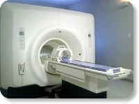
(Signa VH/i 3.0T)
With GE Healthcare
leading-edge technology in ultra-high-field imaging. The 3 T VH/i provides a platform for advanced applications in radiology, cardiology, psychology and psychiatry. Real-time image processing lets you acquire multislice whole brain images and map brain functions for research or surgical planning. And the 3 T Signa VH/i is flexible enough to provide clinicians with high performance they require. It can provide not only outstanding features in brain scanning and neuro-system research, but also a wide range of use in scanning breasts, extremities, the spine and the cardiovascular systems.
Device Information and Specification CLINICAL APPLICATION Whole body
T/R quadrature head, T/R quadrature body, T/R phased array extremity (opt) SE, IR, 2D/3D GRE, FGRE, RF-spoiled GRE, FSE, Angiography: 2D/3D TOF, 2D/3D phase contrast vascular IMAGING MODES Single, multislice, volume study, fast scan, multi slab, cine, localizer 100 Images/sec with Reflex100 MULTISLICE 100 Images/sec with Reflex100 2D 0.5-100mm in 0.1mm incremental 128x512 steps 32 phase encode H*W*D 260cm x 238cm x 265cm POWER REQUIREMENTS 480 or 380/415, 3 phase ||
COOLING SYSTEM TYPE Closed-loop water-cooled grad. Less than 0.14 L/hr liquid He | |  | |
• View the DATABASE results for 'Signa 3.0T™' (2).
| | | | |
|  |  | Searchterm 'Superconducting' was also found in the following services: | | | | |
|  |  |
| |
|

From GE Healthcare;
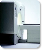
GE's Signa Contour/i system uses the innovations like K4 technology and real-time interactive imaging.
This compact magnet with wide-flare gantry obtains high patient comfort with low costs.
Device Information and Specification CLINICAL APPLICATION Whole body Head and body coil standard; all other coils optional; open architecture makes system compatible with a wide selection of coils Standard: SE, IR, 2D/3D GRE and SPGR, Angiography;; 2D/3D TOF, 2D/3D Phase Contrast;; 2D/3D FSE, 2D/3D FGRE and FSPGR, SSFP, FLAIR, optional: EPI, 2D/3D Fiesta, FGRET, Spiral2D 0.8 mm to 20 mm; 3D 0.1 mm to 5 mm 128x512 steps 32 phase encode POWER REQUIREMENTS 480 or 380/415 V STRENGTH SmartSpeed 23 mT/m, HiSpeed Plus 33 mT/m | |  | | | |
|  | |  |  |  |
| |
|

From GE Healthcare;
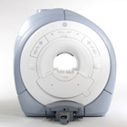
GE Healthcare has added the Signa HDe 1.5T™, a compact MRI device at an affordable price to its family of MRI products. It has a single electronic cabinet that can be positioned inside the scanner room rather than
in a separate equipment room. The Signa HDe 1.5T can be installed in the same physical location as 0.5T MRI systems with minimal construction costs. According to GE, the installation has been simplified to last only 7 days and has a 30 percent smaller footprint than a typical 1.5T system.
The 1.5T Signa™ HDe MRI system is substantially equivalent to the currently marketed GE 1.5T machines. The data acquisition system supports 1, 4, 8 independent receive channels and multiple independent coil elements per channel during a single acquisition series. The gradient specifications of HDe are lower than other GE Signa 1.5T MRI systems, but it can support clinical applications in cardiac and spectroscopy imaging.
Device Information and Specification CLINICAL APPLICATION Whole body CONFIGURATION Compact short bore 2D 0.7 mm to 20 mm; 3D 0.1 mm to 5 mm 128x512 steps 32 phase encode POWER REQUIREMENTS 480 or 380/415 less than 0.03 L/hr liquid helium | |  | |
• View the NEWS results for 'Signa HDe 1.5T™' (1).
| | | | |  Further Reading: Further Reading: | Basics:
|
|
| |
|  | |  |  |
|  | |
|  | | |
|
| |
 | Look
Ups |
| |