Magnetic Resonance - Technology Information Portal Welcome to MRI Technology
Info
  Sheets

Out-
      side


 'Susceptibility' 
SEARCH FOR    
 
  2 3 5 A B C D E F G H I J K L M N O P Q R S T U V W X Y Z
Result : Searchterm 'Susceptibility' found in 3 terms [] and 43 definitions []
previous     41 - 45 (of 46)     next
Result Pages : [1]  [2 3 4 5 6 7 8 9 10]
Searchterm 'Susceptibility' was also found in the following services: 
spacer
News  (3)  Resources  (1)  Forum  (6)  
 
Reticuloendothelial Contrast AgentsInfoSheet: - Contrast Agents - 
Intro, Overview, 
Characteristics, 
Types of, 
etc.MRI Resource Directory:
 - Contrast Agents -
 
Liver imaging with gadolinium contrast enhanced MRI is sometimes not sufficient for a reliable diagnosis of liver lesions. For this reasons, special liver Contrast agents that are targeted to the reticuloendothelial system (RES), have been developed to improve both detection and characterization of liver and spleen lesions. Reticuloendothelial Contrast Agents, as e.g. superparamagnetic iron oxides (SPIO), are taken up by healthy liver tissue but not tumors.
These RES targeted contrast agents provide a prolonged imaging window and enough time for high spatial resolution or multiple breath hold images. Reticuloendothelial contrast agents have an increased sensitivity for the detection of small liver lesions (e.g., metastases), compared with gadolinium enhanced MRI and spiral CT. At higher field strengths with an increased signal to noise ratio the susceptibility effect with iron oxide particles may be enhanced.
Other new agents (Gadobenate Dimeglumine, Gadoxetic Acid) have both an initial extracellular circulation and a delayed liver-specific uptake. Since a considerable part of these contrast agents is excreted in the bile, functional biliary imaging can diagnose biliary anomalies, postoperative bile leaks, and anastomotic strictures. Other agents, such as liposomes (with encapsulated Gd-DTPA) or DOTA complexes are in different development stages.

See also Hepatobiliary Contrast Agents, Gadolinium Oxide, Superparamagnetic Iron Oxide and Liposomes.
spacer
MRI Resources 
Spectroscopy - Online Books - Spectroscopy pool - Implant and Prosthesis pool - Collections - MR Guided Interventions
 
Spin EchoForum -
related threads
 
(SE) The Reappearance of the MR signal after the FID has apparently died away, as a result of the effective reversal (rephasing) of the dephasing spins by techniques such as specific RF pulse sequences or pairs of field gradient pulses, applied in time shorter than or on the order of T2. Proper selection of the TE time of the pulse sequence can help to control the amount of T1 or T2 contrast present in the image. Pulse sequences of the spin echo type, usually employs a 90° pulse, followed by one or more 180° pulses to eliminate field inhomogeneity and chemical shift effects at the echo. Caused by this 180° refocusing pulse, spin echo or fast spin echo (FSE, TSE) sequences are more robust against e.g. susceptibility artifacts than sequences of the gradient echo type.
spacer

• View the DATABASE results for 'Spin Echo' (96).Open this link in a new window


• View the NEWS results for 'Spin Echo' (1).Open this link in a new window.
 
Further Reading:
  Basics:
Spin echoes, CPMG and T2 relaxation - Introductory NMR & MRI from Magritek
2013   by www.azom.com    
  News & More:
EVALUATION OF HUMAN STROKE BY MR IMAGING
2000
MRI Resources 
Examinations - Guidance - Functional MRI - Quality Advice - Pregnancy - Directories
 
Spin Echo SequenceInfoSheet: - Sequences - 
Intro, 
Overview, 
Types of, 
etc.MRI Resource Directory:
 - Sequences -
 
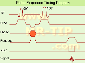
Spin Echo Timing Diagram (SE) The most common pulse sequence used in MR imaging is based of the detection of a spin or Hahn echo. It uses 90° radio frequency pulses to excite the magnetization and one or more 180° pulses to refocus the spins to generate signal echoes named spin echoes (SE).
In the pulse sequence timing diagram, the simplest form of a spin echo sequence is illustrated.
The 90° excitation pulse rotates the longitudinal magnetization (Mz) into the xy-plane and the dephasing of the transverse magnetization (Mxy) starts.
The following application of a 180° refocusing pulse (rotates the magnetization in the x-plane) generates signal echoes. The purpose of the 180° pulse is to rephase the spins, causing them to regain coherence and thereby to recover transverse magnetization, producing a spin echo.
The recovery of the z-magnetization occurs with the T1 relaxation time and typically at a much slower rate than the T2-decay, because in general T1 is greater than T2 for living tissues and is in the range of 100-2000 ms.
The SE pulse sequence was devised in the early days of NMR days by Carr and Purcell and exists now in many forms: the multi echo pulse sequence using single or multislice acquisition, the fast spin echo (FSE/TSE) pulse sequence, echo planar imaging (EPI) pulse sequence and the gradient and spin echo (GRASE) pulse sequence;; all are basically spin echo sequences.
In the simplest form of SE imaging, the pulse sequence has to be repeated as many times as the image has lines.
Contrast values:
PD weighted: Short TE (20 ms) and long TR.
T1 weighted: Short TE (10-20 ms) and short TR (300-600 ms)
T2 weighted: Long TE (greater than 60 ms) and long TR (greater than 1600 ms)
With spin echo imaging no T2* occurs, caused by the 180° refocusing pulse. For this reason, spin echo sequences are more robust against e.g., susceptibility artifacts than gradient echo sequences.

See also Pulse Sequence Timing Diagram to find a description of the components.
 
Images, Movies, Sliders:
 Shoulder Coronal T1 SE  Open this link in a new window
    

Courtesy of  Robert R. Edelman
 Shoulder Axial T1 SE  Open this link in a new window
 MRI Orbita T1  Open this link in a new window
    
 
spacer

• View the DATABASE results for 'Spin Echo Sequence' (24).Open this link in a new window

 
Further Reading:
  Basics:
Fast Spin Echo(.pdf)
Tuesday, 24 January 2006   by www.81bones.net    
Magnetic resonance imaging
   by www.scholarpedia.org    
FUNDAMENTALS OF MRI: Part I
   by www.e-radiography.net    
  News & More:
New MR sequence helps radiologists more accurately evaluate abnormalities of the uterus and ovaries
Thursday, 23 April 2009   by www.eurekalert.org    
MRI techniques improve pulmonary embolism detection
Monday, 19 March 2012   by medicalxpress.com    
Searchterm 'Susceptibility' was also found in the following services: 
spacer
News  (3)  Resources  (1)  Forum  (6)  
 
Superparamagnetic Contrast AgentsInfoSheet: - Contrast Agents - 
Intro, Overview, 
Characteristics, 
Types of, 
etc.MRI Resource Directory:
 - Contrast Agents -
 
Small particles of ferrite are used as superparamagnetic contrast medium in MR imaging (appearing predominantly dark on MRI). These agents exhibit strong T1 relaxation properties, and due to susceptibility differences to their surroundings also produce a strongly varying local magnetic field, which enhances T2 relaxation to darken the contrast media containing structures.
Superparamagnetic contrast agents are also known by the abbreviation SPIO's (small particle iron oxide or superparamagnetic iron oxide) and USPIO's (ultrasmall particle iron oxide or ultrasmall superparamagnetic iron oxide).
Two types of USPIO will be available on the market as blood pool agents, while SPIO's have been used as darkening contrast agents for liver imaging. As particulate matter they are taken up by the RES. Very small particles of less than 300 nanometers also remain intravascular for a prolonged period of time and thus can serve as blood pool agents.

See also the related poll result: 'The development of contrast agents in MRI is'
spacer

• View the DATABASE results for 'Superparamagnetic Contrast Agents' (12).Open this link in a new window

 
Further Reading:
  News & More:
Optimized Labelling of Human Monocytes with Iron Oxide MR Contrast Agents
Sunday, 30 November 2003   by rsna2003.rsna.org    
Poly (dopamine) coated superparamagnetic iron oxide nanocluster for noninvasive labeling, tracking, and targeted delivery of adipose tissue-derived stem cells
Tuesday, 5 January 2016   by www.nature.com    
MRI Resources 
General - Open Directory Project - Knee MRI - NMR - Diffusion Weighted Imaging - Corporations
 
Turbo Gradient Spin EchoInfoSheet: - Sequences - 
Intro, 
Overview, 
Types of, 
etc.MRI Resource Directory:
 - Sequences -
 
(TGSE / TurboGSE) A sequence with a combination of Gradient and Spin Echo Imaging. Additional gradient echoes are generated before and after each spin echo. The spin echoes are allocated to the center of the raw data matrix to give pure T2 contrast. The gradient echoes primarily determine the image resolution. If multiple image lines are obtained during a single echo, the imaging pulse sequence type is a TGSE pulse sequence. This sequence is very fast, fat is darker and more sensitive to susceptibility effects. Also called GRASE.
spacer
MRI Resources 
Jobs - Pregnancy - Process Analysis - Chemistry - Mobile MRI - Manufacturers
 
previous      41 - 45 (of 46)     next
Result Pages : [1]  [2 3 4 5 6 7 8 9 10]
 Random Page
 
Share This Page
FacebookTwitterLinkedIn

MR-TIP    
Community   
User
Pass
Forgot your UserID/Password ?    


New acceleration techniques will :
reduce scan times 
cause artifacts 
increase expenses 
be useful if you have a lot of experience 
doesn't do much 
never heard of 

Look
      Ups


MR-TIP.com uses cookies! By browsing MR-TIP.com, you agree to our use of cookies.

Magnetic Resonance - Technology Information Portal
Member of SoftWays' Medical Imaging Group - MR-TIP • Radiology-TIP • Medical-Ultrasound-Imaging • 
Copyright © 2003 - 2024 SoftWays. All rights reserved. [ 21 November 2024]
Terms of Use | Privacy Policy | Advertising
 [last update: 2024-02-26 03:41:00]