Magnetic Resonance - Technology Information Portal Welcome to MRI Technology
Info
  Sheets

Out-
      side


 'T1 Weighted Image' 
SEARCH FOR    
 
  2 3 5 A B C D E F G H I J K L M N O P Q R S T U V W X Y Z
Result : Searchterm 'T1 Weighted Image' found in 1 term [] and 17 definitions [], (+ 14 Boolean[] results
previous     6 - 10 (of 32)     next
Result Pages : [1]  [2 3 4]  [5 6 7]
Searchterm 'T1 Weighted Image' was also found in the following services: 
spacer
News  (1)  Resources  (1)  Forum  (2)  
 
Fast Low Angle ShotInfoSheet: - Sequences - 
Intro, 
Overview, 
Types of, 
etc.MRI Resource Directory:
 - Sequences -
 
(FLASH) A fast sequence producing signals called gradient echo with low flip angles. FLASH sequences are modifications, which incorporate or remove the effects of transverse coherence respectively.
FLASH uses a semi-random spoiler gradient after each echo to spoil the steady state (to destroy any remaining transverse magnetization) by causing a spatially dependent phase shift. The transverse steady state is spoiled but the longitudinal steady state depends on the T1 values and the flip angle. Extremely short TR times are possible, as a result the sequence provides a mechanism for gaining extremely high T1 contrast by imaging with TR times as brief as 20 to 30 msec while retaining reasonable signal levels. It is important to keep the TE as short as possible to suppress susceptibility artifacts.
The T1 contrast depends on the TR as well as on flip angle, with short TE.
Small flip angles and short TR results in proton density, and long TR in T2* weighting.
With large flip angles and short TR result T1 weighted images.

TR and flip angle adjustment:

TR 3000 ms, Flip Angle 90°
TR 1500 ms, Flip Angle 45°
TR 700 ms, Flip Angle 25°
TR 125 ms, Flip Angle 10°

The apparent ability to trade TR against flip angle for purposes of contrast and the variation in SNR as the scan time (TR) is reduced.

See also Gradient Echo Sequence.
 
Images, Movies, Sliders:
 Fetus (Brain) and Dermoid in Mother  Open this link in a new window
      

Courtesy of  Robert R. Edelman

 
spacer
 
• Related Searches:
    • Gradient Echo Sequence
    • Spoiled Gradient Echo Sequence
    • Liver Imaging
    • T1 Weighted Image
    • Ultrafast Gradient Echo Sequence
 
Further Reading:
  News & More:
Motion Compensation in MR Imaging
   by ccn.ucla.edu    
Turbo-FLASH Based Arterial Spin Labeled Perfusion MRI at 7 T
Thursday, 20 June 2013   by www.plosone.org    
Usefulness of MR Imaging for Diseases of the Small Intestine: Comparison with CT
2000   by www.ncbi.nlm.nih.gov    
MRI Resources 
Education pool - Liver Imaging - IR - Cochlear Implant - Journals - Safety Training
 
Fast Spin EchoForum -
related threadsInfoSheet: - Sequences - 
Intro, 
Overview, 
Types of, 
etc.
 
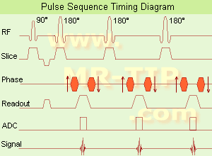
Fast Spin Echo Diagram (FSE) In the pulse sequence timing diagram, a fast spin echo sequence with an echo train length of 3 is illustrated. This sequence is characterized by a series of rapidly applied 180° rephasing pulses and multiple echoes, changing the phase encoding gradient for each echo.
The echo time TE may vary from echo to echo in the echo train. The echoes in the center of the K-space (in the case of linear k-space acquisition) mainly produce the type of image contrast, whereas the periphery of K-space determines the spatial resolution. For example, in the middle of K-space the late echoes of T2 weighted images are encoded. T1 or PD contrast is produced from the early echoes.
The benefit of this technique is that the scan duration with, e.g. a turbo spin echo turbo factor / echo train length of 9, is one ninth of the time. In T1 weighted and proton density weighted sequences, there is a limit to how large the ETL can be (e.g. a usual ETL for T1 weighted images is between 3 and 7). The use of large echo train lengths with short TE results in blurring and loss of contrast. For this reason, T2 weighted imaging profits most from this technique.
In T2 weighted FSE images, both water and fat are hyperintense. This is because the succession of 180° RF pulses reduces the spin spin interactions in fat and increases its T2 decay time. Fast spin echo (FSE) sequences have replaced conventional T2 weighted spin echo sequences for most clinical applications. Fast spin echo allows reduced acquisition times and enables T2 weighted breath hold imaging, e.g. for applications in the upper abdomen.
In case of the acquisition of 2 echoes this type of a sequence is named double fast spin echo / dual echo sequence, the first echo is usually density and the second echo is T2 weighted image. Fast spin echo images are more T2 weighted, which makes it difficult to obtain true proton density weighted images. For dual echo imaging with density weighting, the TR should be kept between 2000 - 2400 msec with a short ETL (e.g., 4).
Other terms for this technique are:
Turbo Spin Echo
Rapid Imaging Spin Echo,
Rapid Spin Echo,
Rapid Acquisition Spin Echo,
Rapid Acquisition with Refocused Echoes
 
Images, Movies, Sliders:
 Lumbar Spine T2 FSE Sagittal  Open this link in a new window
    

Courtesy of  Robert R. Edelman
 MRI - Anatomic Imaging of the Foot  Open this link in a new window
    
SlidersSliders Overview

 Lumbar Spine T2 FSE Axial  Open this link in a new window
    

Courtesy of  Robert R. Edelman
 
spacer

• View the DATABASE results for 'Fast Spin Echo' (31).Open this link in a new window

 
Further Reading:
  Basics:
MYELIN-SELECTIVE MRI: PULSE SEQUENCE DESIGN AND OPTIMIZATION
   by www.imaging.robarts.ca    
Advances in Magnetic Resonance Neuroimaging
Friday, 27 February 2009   by www.ncbi.nlm.nih.gov    
  News & More:
New MR sequence helps radiologists more accurately evaluate abnormalities of the uterus and ovaries
Thursday, 23 April 2009   by www.eurekalert.org    
Spin echoes, CPMG and T2 relaxation - Introductory NMR & MRI from Magritek
2013   by www.azom.com    
MRI Resources 
PACS - Jobs - Absorption and Emission - Safety Training - - Veterinary MRI
 
Gadopentetate DimeglumineInfoSheet: - Contrast Agents - 
Intro, Overview, 
Characteristics, 
Types of, 
etc.MRI Resource Directory:
 - Contrast Agents -
 
Short name: Gd-DTPA, generic name: Gadopentetate dimeglumine, chemical compound: Gadolinium-diethylenetriaminepentaacetic acid
Gadopentetate dimeglumine was introduced in 1981, as the first paramagnetic MRI contrast agent (ionic). The Gd-induced dipole dipole interactions lead to shortening of T1, which results in contrast enhancement on T1 weighted images. The used metal ion Gd3+ (gadolinium) is toxic, and therefore bound in the renally excreted DTPA chelate, a very stable complex. The Gd-complex also induce susceptibility effects, as a result of the magnetic field gradient between the contrast agent in the blood vessels and the surrounding tissue, that lead to shortening of T2 or T2*.
Following intravenous administration, the compound is distributed rapidly in the extracellular space and is eliminated unchanged by glomerular filtration via the kidneys. Up to 6 hours, post injection an average of 83% of the dose is eliminated renal.

See also Magnevist®, Gadolinium and Contrast Agents.
spacer

• View the DATABASE results for 'Gadopentetate Dimeglumine' (5).Open this link in a new window

 
Further Reading:
  Basics:
Magnevist Package Insert
2000
Gadopentetic acid
   by en.wikipedia.org    
  News & More:
EMA's final opinion confirms restrictions on use of linear gadolinium agents in body scans
Friday, 21 July 2017   by www.ema.europa.eu    
Searchterm 'T1 Weighted Image' was also found in the following services: 
spacer
News  (1)  Resources  (1)  Forum  (2)  
 
Gastrointestinal Paramagnetic Contrast AgentsInfoSheet: - Contrast Agents - 
Intro, Overview, 
Characteristics, 
Types of, 
etc.MRI Resource Directory:
 - Contrast Agents -
 
Paramagnetic substances, for example Gd-DTPA solutions, are used as MRI oral contrast agents in gastrointestinal imaging to depict the lumen of the digestive organs. Different Gd-DTPA solutions or zeolites containing gadolinium can be used e.g., for diagnosis of delayed gastric emptying, diagnosis of Crohn's disease etc.
Low concentrations of gastrointestinal paramagnetic contrast agents cause a reduction in T1 relaxation time; consequently, these agents act on T1 weighted images by increasing the signal intensity of the bowel lumen. High concentrations cause T2 shortening by decreasing the signal, similar to superparamagnetic iron oxide. Gd-DTPA chelates are unstable at the low pH in the stomach, therefore buffering is necessary for oral use.

See also Gadopentetate Gastrointestinal, Gadolinium Zeolite, Negative Oral Contrast Agents, Gastrointestinal Superparamagnetic Contrast Agents, and Ferric ammonium citrate.
 
Images, Movies, Sliders:
 MR Colonography Gadolinium per Rectum  Open this link in a new window
      

Courtesy of  Robert R. Edelman
 
spacer

• View the DATABASE results for 'Gastrointestinal Paramagnetic Contrast Agents' (5).Open this link in a new window

MRI Resources 
Guidance - Manufacturers - Cochlear Implant - Quality Advice - Anatomy - Chemistry
 
Incoherent Gradient Echo (Gradient Spoiled)InfoSheet: - Sequences - 
Intro, 
Overview, 
Types of, 
etc.
 
The incoherent gradient echo (gradient spoiled) type of sequence uses a continuous shifting of the RF pulse to spoil the remaining transverse magnetization. The transverse magnetization is destroyed by a magnetic field gradient. This results in a T1 weighted image. Spoiling can be accomplished by RF or a gradient.
Gradient spoiling occurs after each echo by using strong gradients in the slice-select direction after the frequency encoding and before the next RF pulse. Because spins in different locations in the magnet thereby experience a variety of magnetic field strengths, they will precess at differing frequencies; as a consequence they will quickly become dephased. Magnetic field gradients are not very efficient at spoiling the transverse steady state. To be effective, the spins must be forced to precess far enough to become phased randomly with respect to the RF excitation pulse. In clinical MRI machines, the field gradients are set up in such a way that they increase and decrease relative to the center of the magnet; the magnetic field at the magnet 'isocenter' does not change.
The T1 weighting increases with the flip angle and the T2* weighting increases with echo time (TE). Typical repetition time (TR) are 30-500 ms and TE less than 15 ms.

See also Ernst Angle.
spacer
MRI Resources 
Brain MRI - Pregnancy - NMR - Mobile MRI Rental - PACS - Cochlear Implant
 
previous      6 - 10 (of 32)     next
Result Pages : [1]  [2 3 4]  [5 6 7]
 Random Page
 
Share This Page
FacebookTwitterLinkedIn

MR-TIP    
Community   
User
Pass
Forgot your UserID/Password ?    


Next big thing in MRI will be :
AI 
remote operator 
personalized protocols 
helium-free 
molecular MRI 
portable MRI 

Look
      Ups


MR-TIP.com uses cookies! By browsing MR-TIP.com, you agree to our use of cookies.

Magnetic Resonance - Technology Information Portal
Member of SoftWays' Medical Imaging Group - MR-TIP • Radiology-TIP • Medical-Ultrasound-Imaging • 
Copyright © 2003 - 2024 SoftWays. All rights reserved. [ 21 November 2024]
Terms of Use | Privacy Policy | Advertising
 [last update: 2024-02-26 03:41:00]