 | Info
Sheets |
| | | | | | | | | | | | | | | | | | | | | | | | |
 | Out-
side |
| | | | |
|
| |
 |
Dear Guest, Your Attention Please: |
|
|
In the next days, the daily page limit for non-members will
decrease to 20 pages to split up resources in favour of the MR-TIP Community.
Beyond this limitation, most resources will still be available for everyone.
If you want to join the MR-TIP Community, please register here.
|
|
| | | |  | Searchterm 'Ultra' was also found in the following services: | | | | |
|  |  |
| |
|

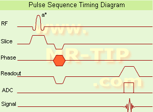
(GRE - sequence) A gradient echo is generated by using a pair of bipolar gradient pulses. In the pulse sequence timing diagram, the basic gradient echo sequence is illustrated. There is no refocusing 180° pulse and the data are sampled during a gradient echo, which is achieved by dephasing the spins with a negatively pulsed gradient before they are rephased by an opposite gradient with opposite polarity to generate the echo.
See also the Pulse Sequence Timing Diagram. There you will find a description of the components.
The excitation pulse is termed the alpha pulse α. It tilts the magnetization by a flip angle α, which is typically between 0° and 90°. With a small flip angle there is a reduction in the value of transverse magnetization that will affect subsequent RF pulses.
The flip angle can also be slowly increased during data acquisition (variable flip angle: tilt optimized nonsaturation excitation).
The data are not acquired in a steady state, where z-magnetization recovery and destruction by ad-pulses are balanced.
However, the z-magnetization is used up by tilting a little more of the remaining z-magnetization into the xy-plane for each acquired imaging line.
Gradient echo imaging is typically accomplished by examining the FID, whereas the read gradient is turned on for localization of the signal in the readout direction. T2* is the characteristic decay time constant associated with the FID. The contrast and signal generated by a gradient echo depend on the size of the longitudinal magnetization and the flip angle.
When α = 90° the sequence is identical to the so-called partial saturation or saturation recovery pulse sequence.
In standard GRE imaging, this basic pulse sequence is repeated as many times as image lines have to be acquired.
Additional gradients or radio frequency pulses are introduced with the aim to spoil to refocus the xy-magnetization at the moment when the spin system is subject to the next α pulse.
As a result of the short repetition time, the z-magnetization cannot fully recover and after a few initial α pulses there is an equilibrium established between z-magnetization recovery and z-magnetization reduction due to the α pulses.
Gradient echoes have a lower SAR, are more sensitive to field inhomogeneities and have a reduced crosstalk, so that a small or no slice gap can be used.
In or out of phase imaging depending on the selected TE (and field strength of the magnet) is possible.
As the flip angle is decreased, T1 weighting can be maintained by reducing the TR.
T2* weighting can be minimized by keeping the TE as short as possible, but pure T2 weighting is not possible.
By using a reduced flip angle, some of the magnetization value remains longitudinal (less time needed to achieve full recovery) and for a certain T1 and TR, there exist one flip angle that will give the most signal, known as the "Ernst angle".
Contrast values:
PD weighted: Small flip angle (no T1), long TR (no T1) and short TE (no T2*)
T1 weighted: Large flip angle (70°), short TR (less than 50ms) and short TE
T2* weighted: Small flip angle, some longer TR (100 ms) and long TE (20 ms)
Classification of GRE sequences can be made into four categories:
See also Gradient Recalled Echo Sequence, Spoiled Gradient Echo Sequence, Refocused Gradient Echo Sequence, Ultrafast Gradient Echo Sequence.
| | | |  | | | | | | | | |  Further Reading: Further Reading: | | Basics:
|
|
News & More:
| |
| |
|  | |  |  |  |
| |
|

Founded in 1991, ImaRx Pharmaceutical Corp. designs, develops and markets pharmaceuticals for medical imaging ( MRI, ultrasound and computed tomography) for the radiological imaging industry.
ImaRx Pharmaceutical Corp., announced 1999 that it has been acquired by E.I DuPont de Nemours and Co., Inc.. The terms of the acquisition provide a royalty-free licensing arrangement with a newly-formed company, ImaRx LLC ("LLC"), to pursue and develop new products and technologies for drug and gene delivery independent from DuPont.
Yamanouchi Pharmaceutical Co. Ltd., ImaRx' licensee for Asian territories for this product, will continue to develop the product in Asia as DuPont's licensee. ImaRx LLC will have ownership of all other targeted and therapeutic products previously owned by ImaRx, including imaging products outside of diagnostic ultrasound imaging and two other imaging products, SonoRx® and LumenHance®, which are both FDA approved and licensed to BRACCO Diagnostics.
MRI Contrast Agents:
Contact Information
MAIL
ImaRx LLC
1635 East 18th Street
Tucson AZ 85719-6803
USA
| |  | | | |
|  | |  |  |  |
| |
|
From Philips Medical Systems;

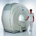
Philips Infinion 1.5 T is designed to maximize the efficiency and quality of patient care. Developed with the patient in mind, the Infinion is the shortest and most open 1.5T scanner available. The unique ' ultra short' 1.4 m magnet assures patient comfort and acceptance without compromising image quality and clinical performance.
Device Information and Specification
CLINICAL APPLICATION
Whole body
CONFIGURATION
Ultra short bore
Head, head / neck, integrated C-spine, L/T spine array, small large GP coils, body flex array, torso pelvis array, breast array, endocavitary, shoulder array, lower extremity, hand / wrist, cardiac, PV array
SE, TSE, SS TSE, EPI, IR, STIR, FLAIR, FFE, TFE, T1 TFE, T2 TFE, Presat, Fatsat, MTC, Diff-opt., Angiography: PCA, MCA, TOF
IMAGING MODES
Single slice, single volume, multi slice, multi volume
80 images/sec std.; up to320 opt.@256
H*W*D
233 (lead fitted) x 198 x 140 cm
POWER REQUIREMENTS
400/480 V
COOLING SYSTEM TYPE
Closed loop, chilled water
| |  | |
• View the DATABASE results for 'Infinion 1.5T™' (2).
| | | | |
|  |  | Searchterm 'Ultra' was also found in the following services: | | | | |
|  |  |
| |
|
Contrast agent with a preferential intracellular distribution.
Intracellular agents (such as manganese derivatives and ultrasmall superparamagnetic iron oxide), exhibit a flow- and metabolism-dependent uptake. These properties may allow delayed imaging, similar to isotopic methods.
Phospholipid liposomes are rapidly sequestered by the cells in the reticuloendothelial system (RES), primarily in the liver. For imaging of the liver, liposomes may be labeled with MR contrast medium, both positive (T1-shortening) paramagnetic media, and negative (T2-shortening) superparamagnetic media.
Several other nonliposome MR contrast media are also taken up by the RES, e.g.:
Other MR contrast agents accumulate selectively in the hepatocytes, e.g.:
| |  | |
• View the DATABASE results for 'Intracellular Contrast Agents' (3).
| | | | |  Further Reading: Further Reading: | News & More:
|
|
| |
|  | |  |  |  |
| |
|
The MRI device is located within a specially shielded room ( Faraday cage) to avoid outside interference, caused by the use of radio waves very close in frequency to those of ordinary FM radio stations.
The MRI procedure can easily be performed through clothing and bones, but attention must be paid to ferromagnetic items, because they will be attracted from the magnetic field. A hospital gown is appropriate, or the patient should wear clothing without metal fasteners and remove any metallic objects like hairpins, jewelry, eyeglasses, clocks, hearing aids, any removable dental work, lighters, coins etc., not only for MRI safety reasons.
Metal in or around the scanned area can also cause errors in the reconstructed images ( artifacts). Because the strong magnetic field can displace, or disrupt metallic objects, people with an implanted active device like a cardiac pacemaker cannot be scanned under normal circumstances and should not enter the MRI area.
The MRI machine can look like a short tunnel or has an open MRI design and the magnet does not completely surround the patient. Usually the patient lies on a comfortable motorized table, which slides into the scanner, depending on the MRI device, patients may be also able to sit up. If a contrast agent is to be administered, intravenous access will be placed. A technologist will operate the MRI machine and observe the patient during the examination from an adjacent room. Several sets of images are usually required, each taking some minutes. A typical MRI scan includes three to nine imaging sequences and may take up to one hour. Improved MRI devices with powerful magnets, newer software, and advanced sequences may complete the process in less time and better image quality.
Before and after the most MRI procedures no special preparation, diet, reduced activity, and extra medication is necessary. The magnetic field and radio waves are not felt and no pain is to expect.
Movement can blur MRI images and cause certain artifacts. A possible problem is the claustrophobia that some patients experience from being inside a tunnel-like scanner. If someone is very anxious or has difficulty to lie still, a sedative agent may be given. Earplugs and/or headphones are usually given to the patient to reduce the loud acoustic noise, which the machine produces during normal operation. A technologist observes the patient during the test. Some MRI scanners are equipped with televisions and music to help the examination time pass.
MRI is not a cheap examination, however cost effective by eliminating the need for invasive radiographic procedures, biopsies, and exploratory surgery. MRI scans can also save money while minimizing patient risk and discomfort. For example, MRI can reduce the need for X-ray angiography and myelography, and can eliminate unnecessary diagnostic procedures that miss occult disease. See also Magnetic Resonance Imaging MRI, Medical Imaging, Cervical Spine MRI, Claustrophobia, MRI Risks and Pregnancy.
For Ultrasound Imaging (USI) see Ultrasound Imaging Procedures at Medical-Ultrasound-Imaging.com.
See also the related poll result: ' MRI will have replaced 50% of x-ray exams by' | | | |  | |
• View the DATABASE results for 'MRI Procedure' (11).
| | |
• View the NEWS results for 'MRI Procedure' (6).
| | | | |  Further Reading: Further Reading: | News & More:
|
|
| |
|  | |  |  |
|  | |
|  | | |
|
| |
 | Look
Ups |
| |