 | Info
Sheets |
| | | | | | | | | | | | | | | | | | | | | | | | |
 | Out-
side |
| | | | |
|
| | | | |
Result : Searchterm 'Vascular Imaging' found in 2 terms [ ] and 19 definitions [ ] and 19 definitions [ ] ]
| previous 16 - 20 (of 21) nextResult Pages :  [1] [1]  [2 3 4 5] [2 3 4 5] |  | |  | Searchterm 'Vascular Imaging' was also found in the following services: | | | | |
|  |  |
| |
|
| | | | | | | | |
• View the NEWS results for 'Heart MRI' (18).
| | | | |  Further Reading: Further Reading: | | Basics:
|
|
News & More:
| |
| |
|  | |  |  |  |
| |
|

From Philips Medical Systems;
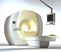
the Intera 3 T high field system, the first with a compact magnet, which is built on the same platform as the 1.5 T, is targeted to high-end neurological, orthopedic and cardiovascular imaging applications with maximum patient comfort and acceptance without compromising image quality and clinical performance. Useable for clinical routine and research.
The Intera systems offer diffusion tensor imaging ( DTI) fiber tracking that measures movement of water in the brain and can therefore detect areas of the brain where normal movement of water is disrupted.
Device Information and Specification
CLINICAL APPLICATION
Whole body
CONFIGURATION
Short bore compact
Standard: head, body, C1, C3; Optional: Small joint, flex-E, flex-R, endocavitary (L and S), dual TMJ, knee, neck, T/L spine, breast; Optional phased array: spine;; Optional SENSE coils: Flex body, flex cardiac, neuro-vascular, head
SE, Modified-SE, IR (T1, T2, PD), STIR, FLAIR, SPIR, FFE, T1-FFE, T2-FFE, Balanced FFE, TFE, Balanced TFE, Dynamic, Keyhole, 3D, Multi Chunk 3D, Multi Stack 3D, K Space Shutter, MTC, TSE, Dual IR, DRIVE, EPI, Cine, 2DMSS, DAVE, Mixed Mode; Angiography: Inflow MRA, TONE, PCA, CE MRA
TR
Min. 1.6 (Master) msec
TE
Min. 0.5 (Master) msec
RapidView Recon. greater than 500 @ 256 Matrix
0.1 mm (Omni), 0.05 mm (Power)
128 x 128, 256 x 256,512 x 512,1024 x 1024 (64 for Bold img)
Variable in 1% increments
Lum.: 120 cd/m2; contrast: 150:1
Variable (op. param. depend.)
POWER REQUIREMENTS
380/400 V
STRENGTH
30 (Master) mT/m
| |  | |
• View the DATABASE results for 'Intera 3.0T™' (2).
| | | | |
|  | |  |  |  |
| |
|
Device Information and Specification
CLINICAL APPLICATION
Whole body
CONFIGURATION
Short bore compact
Standard: Head, body, cardiac, optional phased array: Spine, pediatric, 3rd party connector; Optional SENSE? coils for all applications
SE, Modified-SE, IR (T1, T2, PD), STIR, FLAIR, SPIR, FFE, T1-FFE, T2-FFE, Balanced FFE, TFE, Balanced TFE, Dynamic, Keyhole, 3D, Multi Chunk 3D, Multi Stack 3D, K Space Shutter, MTC, TSE, Dual IR, DRIVE, EPI, Cine, 2DMSS, DAVE, Mixed Mode; Angiography: Inflow MRA, TONE, PCA, CE MRA
128 x 128, 256 x 256,512 x 512,1024 x 1024 (64 for Bold img)
Variable in 1% increments
Lum.: 120 cd/m2; contrast: 150:1
Variable (op. param. depend.)
POWER REQUIREMENTS
380/400 V
| |  | |
• View the DATABASE results for 'Intera Achieva CV™' (2).
| | | | |  Further Reading: Further Reading: | News & More:
|
|
| |
|  |  | Searchterm 'Vascular Imaging' was also found in the following services: | | | | |
|  |  |
| |
|
MRI of the lumbar spine, with its multiplanar 3 dimensional imaging capability, is currently the preferred modality for establishing a diagnosis. MRI scans and magnetic resonance myelography have many advantages compared with computed tomography and/or X-ray myelography in evaluating the lumbar spine. MR imaging scans large areas of the spine without ionizing radiation, is noninvasive, not affected by bone artifacts, provides vascular imaging capability, and makes use of safer contrast agents ( gadolinium chelate).
Due to the high level of tissue contrast resolution, nerves and discs are clearly visible. MRI is excellent for detecting degenerative disease in the spine. Lumbar spine MRI accurately shows disc disease (prolapsed disc or slipped disc), the level at which disc disease occurs, and if a disc is compressing spinal nerves. Lumbar spine MRI depicts soft tissues, including the cauda equina, spinal cord, ligaments, epidural fat, subarachnoid space, and intervertebral discs. Loss of epidural fat on T1 weighted images, loss of cerebrospinal fluid signal around the dural sac on T2 weighted images and degenerative disc disease are common features of lumbar stenosis.
Common indications for MRI of the lumbar spine:
•
Neurologic deficits, evidence of radiculopathy, acute spinal cord compression (e.g., sudden bowel/bladder disturbance)
•
Suspected systemic disorders (primary tumors, drop metastases, osteomyelitis)
•
Postoperative evaluation of lumbar spine: disk vs. scar
•
Localized back pain with no radiculopathy (leg pain)
Lumbar spine imaging requires a special spine coil. often used whole spine array coils have the advantage that patients do not need other positioning if also upper parts of the spine should be scanned. Sagittal T1 and T2 weighted FSE sequences are the standard views. With multi angle oblique techniques individually oriented transverse images of each intervertebral disc at different angles can be obtained.
See also the related poll result: ' MRI will have replaced 50% of x-ray exams by' | | | |  | |
• View the DATABASE results for 'Lumbar Spine MRI' (6).
| | | | |  Further Reading: Further Reading: | Basics:
|
|
News & More:
| |
| |
|  | |  |  |  |
| |
|
The definition of a scan is to form an image or an electronic representation. The MRI scan uses magnetic resonance principles to produce extremely detailed pictures of the body tissue without the need for X-ray exposure or other damaging forms of radiation.
MRI scans show structures of the different tissues in the body. The tissue that has the least hydrogen atoms (e.g., bones) appears dark, while the tissue with many hydrogen atoms (e.g., fat) looks bright. The MRI pictures of the brain show details and abnormal structures ( brain MRI), for example, tumors, multiple sclerosis lesions, bleedings, or brain tissue that has suffered lack of oxygen after a stroke.
A cardiac MRI scan demonstrates the heart as well as blood vessels ( cardiovascular imaging) and is used to detect heart defects with e.g., changes in the thickness and infarctions of the muscles around the heart. With MRI scans, nearly all kind of body parts can be tested, for example the joints like knee and shoulder, lumbar, thoracic and cervical spine, the pelvis including fetal MRI, and the soft parts of the body such as the liver, kidneys, and spleen.
The MRI procedure includes three to nine imaging sequences and may take up to one hour. See also Lumbar Spine MRI, MRI Safety and Open MRI. | | | | | | | | | | |
• View the DATABASE results for 'MRI Scan' (31).
| | |
• View the NEWS results for 'MRI Scan' (95).
| | | | |  Further Reading: Further Reading: | Basics:
|
|
News & More:
|  |
A Knee MRI in Half the Time? It's Possible
Thursday, 8 April 2021 by www.diagnosticimaging.com |  |  |
Michigan radiologist warns about 'incidental findings' in full body MRI scans
Wednesday, 4 October 2023 by www.wilx.com |  |  |
ACCELERATING MRI SCANS WITH ARTIFICIAL INTELLIGENCE
Friday, 28 August 2020 by www.analyticsinsight.net |  |  |
Radiographer's Lego Open MRI Product Idea Reaches New Milestone
Monday, 11 November 2019 by www.itnonline.com |  |  |
Why we need erasable MRI scans
Wednesday, 25 April 2018 by phys.org |  |  |
MRI as accurate as CT for Crohn's disease detection, management
Tuesday, 6 June 2017 by www.healthimaging.com |  |  |
MRI scans predict patients' ability to fight the spread of cancer
Tuesday, 12 December 2017 by eurekalert.org |  |  |
Audio/Video System helps patients relax during MRI scans
Monday, 8 December 2014 by news.thomasnet.com |  |  |
MRI scans could be a 'game-changer' in prostate cancer testing
Tuesday, 5 August 2014 by www.abc.net.au |  |  |
7-Tesla MRI scanner allows even more accurate diagnosis of breast cancer
Thursday, 6 March 2014 by www.healthcanal.com |
|
| |
|  | |  |  |
|  | | |
|
| |
 | Look
Ups |
| |