 | Info
Sheets |
| | | | | | | | | | | | | | | | | | | | | | | | |
 | Out-
side |
| | | | |
|
| | | | |
Result : Searchterm 'Vascular Imaging' found in 2 terms [ ] and 19 definitions [ ] and 19 definitions [ ] ]
| 1 - 5 (of 21) nextResult Pages :  [1] [1]  [2 3 4 5] [2 3 4 5] |  | |  | Searchterm 'Vascular Imaging' was also found in the following services: | | | | |
|  |  |
| |
|
Cardiovascular MR imaging includes the complete anatomical display of the heart with CINE imaging of all phases of the heartbeat. Ultrafast techniques make breath hold three-dimensional coverage of the heart in different cardiac axes feasible. Cardiac MRI provides reliable anatomical and functional assessment of the heart and evaluation of myocardial viability and coronary artery disease by a noninvasive diagnostic imaging technique.
Cardiovascular MRI offers potential advantages over radioisotopic techniques because it provides superior spatial resolution, does not use ionizing radiation, has no imaging orientations constraints and contrast resolution better than echocardiography. It also offers direct visualization and characterization of atherosclerotic plaques and diseased vessel walls and surrounding tissues in cardiovascular research.
MRI perfusion approaches measure the alteration of regional myocardial magnetic properties after the intravenous injection of contrast agents and assess the extent of injury after a myocardial infarction and the presence of myocardial viability with a technique based on late enhancement. Extracellular MRI contrast agents, like Gd-DTPA, accumulate only in irreversibly damaged myocardium after a time period of at least 10 minutes.
This type of patients may also have an implanted cardiac stent, bypass or a cardiac pacemaker and special caution should be observed on the MRI safety and the contraindications. While a number of coronary stents have been tested and reported to be MRI compatible, coronary stents must be assessed on an individual basis, with the medical team weighing the risks and benefits of the MRI procedure.
Cardiac MRI overview:
•
Calculation of ventricular volume, myocardial mass and wall thickness
•
Description of a stenosis or aneurysma
•
Anatomical display of the heart, vessels and the surrounding tissue
Cardiovascular MRI has become one of the most effective noninvasive imaging techniques for almost all groups of heart and vascular disease. | | | |  | | | | • Share the entry 'Cardiovascular Imaging':    | | |
• View the NEWS results for 'Cardiovascular Imaging' (6).
| | | | |  Further Reading: Further Reading: | | Basics:
|
|
News & More:
| |
| |
|  | |  |  |  |
| |
|
The use of gas as a contrast medium has significant potential to avoid limitations of conventional contrast agents. Gases can transit smaller vascular conduits and can be injected through smaller and less traumatic access systems than liquids. Highly soluble gases (such as CO2) can be imaged as a bolus. Blood is displaced by the gas, with the result of negative image contrast.
Because gases are compressible, standard liquid injectors
cannot be used. The design for a gasinjector should have the option for individual adaptation of blood flow rate, vessel diameter, pulse pressure, and heart rate. | |  | | | |  Further Reading: Further Reading: | News & More:
|
|
| |
|  | |  |  |  |
| |
|

From Toshiba America Medical Systems Inc.;
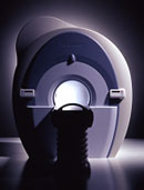
With its high-field strength, the Vantage™ delivers the clinical capabilities and image quality expected by cardiologists, while simultaneously offering patients a more comfortable and non-invasive option, said Dane Peshe, director, MRI Business Unit, Toshiba America Medical Systems. Vantage™ supports a full complement of cardiovascular imaging studies, ranging from stroke evaluation to peripheral vascular imaging. Additionally, the ultra short bore design offers patients a greater feeling of openness that reduces claustrophobic sensations, while Toshiba's exclusive, patented Pianissimo™ technology reduces scan noise by as much as 90 percent for a more pleasant experience.'
Device Information and Specification CLINICAL APPLICATION Whole body CONFIGURATION Ultra short bore SE, FE, IR, FastSE, FastIR, FastFLAIR, Fast STIR, FastFE, FASE, EPI, SuperFASE; Angiography: 2D(gate/non-gate)/3D TOF, SORS-STC IMAGING MODES Single, multislice, volume study 32-1024, phase;; 64-1024, freq. POWER REQUIREMENTS 380/400/415/440/480 V COOLING SYSTEM TYPE Closed-loop water-cooled Liquid helium: approx. less than 0.05 L/hr Passive, active, auto-active | |  | |
• View the DATABASE results for 'Vantage™' (2).
| | | | |  Further Reading: Further Reading: | Basics:
|
|
| |
|  |  | Searchterm 'Vascular Imaging' was also found in the following services: | | | | |
|  |  |
| |
|
ABLAVAR™ (formerly named Vasovist™) is a blood pool agent for magnetic resonance angiography ( MRA), which opens new medical imaging possibilities in the evaluation of aortoiliac occlusive disease (AIOD) in patients with suspected peripheral vascular disease.
ABLAVAR™ binds reversibly to blood albumin, providing imaging with high spatial resolution up to 1 hour after injection, due to its high relaxivity and to the long lasting increased signal intensity of blood.
As with other contrast media: the possibility of serious or life-threatening anaphylactic or anaphylactoid reactions, including cardiovascular, respiratory and/or cutaneous manifestations, should always be considered.
WARNING:
NEPHROGENIC SYSTEMIC FIBROSIS
Gadolinium-based contrast agents increase the risk for nephrogenic systemic fibrosis (NSF) in patients with acute or chronic severe renal insufficiency (glomerular filtration rate less than 30 mL/min/1.73m 2), or acute renal insufficiency of any severity due to the hepato-renal syndrome or in the perioperative liver transplantation period.
See also Cardiovascular Imaging, Adverse Reaction, Molecular Imaging, and MRI Safety.
Drug Information and Specification
NAME OF COMPOUND
Diphenylcyclohexyl phosphodiester-Gd-DTPA, gadofosveset trisodium, MS-325
T1, predominantly positive enhancement
20-45 mmol-1sec-1, Bo=0,47T
PHARMACOKINETIC
Intravascular
CONCENTRATION
244 mg/mL, 0.25mmol/mL
DOSAGE
0.12 mL/kg, 0.03 mmol/kg
DEVELOPMENT STAGE
FDA approved
DO NOT RELY ON THE INFORMATION PROVIDED HERE, THEY ARE
NOT A SUBSTITUTE FOR THE ACCOMPANYING
PACKAGE INSERT!
Distribution Information
TERRITORY
TRADE NAME
DEVELOPMENT
STAGE
DISTRIBUTOR
USA, Canada, Australia
ABLAVAR™
Approved
| |  | |
• View the DATABASE results for 'ABLAVAR™' (3).
| | |
• View the NEWS results for 'ABLAVAR™' (1).
| | | | |  Further Reading: Further Reading: | Basics:
|
|
News & More:
| |
| |
|  | |  |  |  |
| |
|
With this method irregular RR intervals in cardiac gating during cardiovascular imaging are rejected and then repeated to improve the image quality, whereby the cardiac frequency is used as a basis of the normal heart rate.
The RR interval window determines the percentage variation of the heart rate. Variations of the acquired data outside the window are rejected and not used in the image reconstruction. Also one interval after the arrhytmic beat will be rejected.
Arrhythmia rejection may be inappropriate for patients with certain pathologies, because if the RR interval is constant long, short, long, - all intervals would be rejected. Also a disadvantage is the time consume, but in some cases this function is mandatory, e.g. for diverse retrospective triggered sequences. | |  | | | |  Further Reading: Further Reading: | Basics:
|
|
News & More:
| |
| |
|  | |  |  |
|  | 1 - 5 (of 21) nextResult Pages :  [1] [1]  [2 3 4 5] [2 3 4 5] |
| |
|
| |
 | Look
Ups |
| |