 | Info
Sheets |
| | | | | | | | | | | | | | | | | | | | | | | | |
 | Out-
side |
| | | | |
|
| | | | |
Result : Searchterm 'brain' found in 3 terms [ ] and 54 definitions [ ] and 54 definitions [ ] ]
| previous 16 - 20 (of 57) nextResult Pages :  [1] [1]  [2 3 4 5 6 7 8 9 10 11 12] [2 3 4 5 6 7 8 9 10 11 12] |  | |  | Searchterm 'brain' was also found in the following services: | | | | |
|  |  |
| |
|
(DWI) Magnetic resonance imaging is sensitive to diffusion, because the diffusion of water molecules along a field gradient reduces the MR signal. In areas of lower diffusion the signal loss is less intense and the display from this areas is brighter. The use of a bipolar gradient pulse and suitable pulse sequences permits the acquisition of diffusion weighted images (images in which areas of rapid proton diffusion can be distinguished from areas with slow diffusion).
Based on echo planar imaging, multislice DWI is today a standard for imaging brain infarction. With enhanced gradients, the whole brain can be scanned within seconds. The degree of diffusion weighting correlates with the strength of the diffusion gradients, characterized by the b-value, which is a function of the gradient related parameters: strength, duration, and the period between diffusion gradients.
Certain illnesses show restrictions of diffusion, for example demyelinization and cytotoxic edema. Areas of cerebral infarction have decreased apparent diffusion, which results in increased signal intensity on diffusion weighted MRI scans. DWI has been demonstrated to be more sensitive for the early detection of stroke than standard pulse sequences and is closely related to temperature mapping.
DWIBS is a new diffusion weighted imaging technique for the whole body that produces PET-like images. The DWIBS sequence has been developed with the aim to detect lymph nodes and to differentiate normal and hyperplastic from metastatic lymph nodes. This may be possible caused by alterations in microcirculation and water diffusivity within cancer metastases in lymph nodes.
See also Diffusion Weighted Sequence, Perfusion Imaging, ADC Map, Apparent Diffusion Coefficient, and Diffusion Tensor Imaging. | |  | | | | | | | | |  Further Reading: Further Reading: | | Basics:
|
|
News & More:
| |
| |
|  | |  |  |  |
| |
|
(Dy) The Dysprosium chelates are analogs of the extracellular gadolinium chelates, with substitution of the dysprosium ion for the gadolinium ion and offers advantages in application on first pass studies.
The use of a dysprosium-based contrast agent (Dy-type) instead of a gadolinium-type (Gd-type) is an alternative in cases of a disrupted blood brain barrier. Because of its much smaller T1 enhancement, this contrast agent should give more accurate perfusion calculations in brain MRI. | |  | |
• View the NEWS results for 'Dysprosium' (1).
| | | | |  Further Reading: Further Reading: | Basics:
|
|
| |
|  | |  |  |  |
| |
|

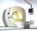
From Philips Medical Systems;
the Intera 3 T high field system, the first with a compact magnet, which is built on the same platform as the 1.5 T, is targeted to high-end neurological, orthopedic and cardiovascular imaging applications with maximum patient comfort and acceptance without compromising image quality and clinical performance. Useable for clinical routine and research.
The Intera systems offer diffusion tensor imaging ( DTI) fiber tracking that measures movement of water in the brain and can therefore detect areas of the brain where normal movement of water is disrupted.
Device Information and Specification
CLINICAL APPLICATION
Whole body
CONFIGURATION
Short bore compact
Standard: head, body, C1, C3; Optional: Small joint, flex-E, flex-R, endocavitary (L and S), dual TMJ, knee, neck, T/L spine, breast; Optional phased array: spine;; Optional SENSE coils: Flex body, flex cardiac, neuro-vascular, head
SE, Modified-SE, IR (T1, T2, PD), STIR, FLAIR, SPIR, FFE, T1-FFE, T2-FFE, Balanced FFE, TFE, Balanced TFE, Dynamic, Keyhole, 3D, Multi Chunk 3D, Multi Stack 3D, K Space Shutter, MTC, TSE, Dual IR, DRIVE, EPI, Cine, 2DMSS, DAVE, Mixed Mode; Angiography: Inflow MRA, TONE, PCA, CE MRA
TR
Min. 1.6 (Master) msec
TE
Min. 0.5 (Master) msec
RapidView Recon. greater than 500 @ 256 Matrix
0.1 mm (Omni), 0.05 mm (Power)
128 x 128, 256 x 256,512 x 512,1024 x 1024 (64 for Bold img)
Variable in 1% increments
Lum.: 120 cd/m2; contrast: 150:1
Variable (op. param. depend.)
POWER REQUIREMENTS
380/400 V
STRENGTH
30 (Master) mT/m
| |  | |
• View the DATABASE results for 'Intera 3.0T™' (2).
| | | | |
|  |  | Searchterm 'brain' was also found in the following services: | | | | |
|  |  |
| |
|
(MSI) The combination of biomagnetic field detection and MR imaging into a merged data set. Most applications of MSI involve the combined use of MRI and measurement of magnetic fields created by electric currents in the brain, so-called magnetoencephalography MEG.
MEG allows calculation of the source of the measured biomagnetic fields, and thereby localization of many regional brain functions, such as mapping of the sensorimotor, auditory and visual cortex and also localization of epileptogenic foci.
The MEG coordinate system is defined by anatomical landmarks, which are easily identified also with MRI, making it possible to align the 3D MEG data with the 3D MR image data. The resulting magnetic source images show the spatial relationships between the functional area provided by MEG and the neighboring anatomy and pathology, both provided by MRI.
Cardiac applications of MSI are also being explored. The electric currents in the myocardium create extrathoracic magnetic fields and the source of these fields may be calculated by the same principles as those used in MEG. Possible cardiac applications include mapping of arrhythmogenic sites prior to ablation therapy. | |  | |
• View the DATABASE results for 'Magnetic Source Imaging' (2).
| | |
• View the NEWS results for 'Magnetic Source Imaging' (2).
| | | | |  Further Reading: Further Reading: | News & More:
|
|
| |
|  | |  |  |  |
| |
|
(MTC) This MRI method increases the contrast by removing a portion of the total signal in tissue. An off resonance radio frequency (RF) pulse saturates macromolecular protons to make them invisible (caused by their ultra-short T2* relaxation times). The MRI signal from semi-solid tissue like brain parenchyma is reduced, and the signal from a more fluid component like blood is retained.
E.g., saturation of broad spectral lines may produce decreases in intensity of lines not directly saturated, through exchange of magnetization between the corresponding states; more closely coupled states will show a greater resulting intensity change.
Magnetization transfer techniques make demyelinated brain or spine lesions (as seen e.g. in multiple sclerosis) better visible on T2 weighted images as well as on gadolinium contrast enhanced T1 weighted images.
Off resonance makes use of a selection gradient during an off resonance MTC pulse. The gradient has a negative offset frequency on the arterial side of the imaging volume (caudally more off resonant and cranially less off resonant). The net effect of this type of pulse is that the arterial blood outside the imaging volume will retain more of its longitudinal magnetization, with more vascular signal when it enters the imaging volume. Off resonance MTC saturates the venous blood, leaving the arterial blood untouched.
On resonance has no effect on the free water pool but will saturate the bound water pool and is the difference in T2 between the pools. Special binomial pulses are transmitted causing the magnetization of the free protons to remain unchanged. The z-magnetization returns to its original value. The spins of the bound pool with a short T2 experience decay, resulting in a destroyed magnetization after the on resonance pulse.
See also Magnetization Transfer. | |  | |
• View the DATABASE results for 'Magnetization Transfer Contrast' (5).
| | | | |  Further Reading: Further Reading: | News & More:
|
|
| |
|  | |  |  |
|  | | |
|
| |
 | Look
Ups |
| |