Magnetic Resonance - Technology Information Portal Welcome to MRI Technology
Info
  Sheets

Out-
      side


 'relaxation' 
SEARCH FOR    
 
  2 3 5 A B C D E F G H I J K L M N O P Q R S T U V W X Y Z
Result : Searchterm 'relaxation' found in 15 terms [] and 75 definitions []
previous     26 - 30 (of 90)     next
Result Pages : [1 2 3]  [4 5 6 7 8 9 10 11 12 13 14 15 16 17 18]
Searchterm 'relaxation' was also found in the following services: 
spacer
News  (8)  Resources  (3)  Forum  (8)  
 
Flow Sensitive Alternating Inversion RecoveryInfoSheet: - Sequences - 
Intro, 
Overview, 
Types of, 
etc.
 
(FAIR) In this sequence 2 inversion recovery images are acquired, one with a nonselective and the other with a slice selective inversion pulse. The z-magnetization in the first sequence is independent of flow. Inflowing spins give z-magnetization from second pulse. A major signal loss in FAIR is the T1 relaxation of tagged blood in transit to the imaging slice. Sharper edges of the inversion pulse give narrow spacing between the inversion edge and the 1st slice because reduced transit time gives lower T1 relaxation induced signal loss. The difference of the images in a consequence contains information proportional to flow (blood partition coefficient). Standard adiabatic inversion RF pulse does not have good slice-profile, because of power/SAR limitation. A c-shaped frequency offset corrected inversion (FOCI) RF pulse can help to increase the signal.
Perfusion imaging, e.g. myocardial, using tissue water as endogenous contrast is suggested.
spacer
MRI Resources 
Research Labs - MRI Technician and Technologist Schools - Manufacturers - Jobs pool - Diffusion Weighted Imaging - Education
 
GadoliniumForum -
related threadsInfoSheet: - Contrast Agents - 
Intro, Overview, 
Characteristics, 
Types of, 
etc.
 
(Gd) Gadolinium is a Lanthanide element that is paramagnetic in its trivalent state.
This paramagnetic substance is used for MR imaging because of the effect of strongly decreasing the T1 relaxation times of the tissues to which gadolinium has access. When injected during magnetic resonance imaging, gadolinium will tend to change signal intensities by shortening the T1 time in its surroundings.
The relaxivity of gadolinium is an important measure of its efficacy, which is dependent on the chemical properties of the complex. The gadolinium ion cannot be used in its chloride, sulfate, or acetate forms because of poor tolerance and low solubility in water in the neutral pH range. Although toxic by itself, gadolinium can be given safely in a chelated form such as DTPA, that still retains much of its strong effect on relaxation times (relaxivity).

See also Dotarem®, Gadovist®, MultiHance®, Omniscan®, OptiMARK®, and Contrast Agents, the info sheet gives an overview and more in-dept information about different types of MRI contrast agents.
 
Images, Movies, Sliders:
 Normal Lung Gd Perfusion MRI  Open this link in a new window
      

Courtesy of  Robert R. Edelman

 Breast MRI Images T1 Pre - Post Contrast  Open this link in a new window
 Delayed Myocardial Contrast Enhancement from Infarct  Open this link in a new window
      

Courtesy of  Robert R. Edelman
 
spacer

• View the DATABASE results for 'Gadolinium' (66).Open this link in a new window


• View the NEWS results for 'Gadolinium' (17).Open this link in a new window.
 
Further Reading:
  Basics:
DNA-SEEKING GADOLINIUM COMPLEXES FOR NEUTRON CAPTURE THERAPY (NCT) (.pdf)
A LANTHANIDE LANTHOLOGY(.pdf)
   by www.phy.davidson.edu    
Gadolinium-Based Agents Safe for MRI Use
Monday, 18 April 2011   by www.renalandurologynews.com    
  News & More:
Stable Spherical Lanthanide Cluster for Magnetic Resonance Imaging Assembly
Wednesday, 26 April 2023   by www.miragenews.com    
Artificial Intelligence Processes Provide Solutions to Gadolinium Retention Concerns
Thursday, 30 January 2020   by www.itnonline.com    
Contrast Agents: Safety Profile
   by www.clinical-mri.com    
Contrast MRIs cause claims, concern, over residual metal in brain
Tuesday, 8 December 2015   by www.afr.com    
Contrast agent linked with brain abnormalities on MRI
Tuesday, 17 December 2013   by www.sciencecodex.com    
FDA Approves First Macrocyclic and Ionic Gadolinium-Contrast Agent for MRI
Friday, 22 March 2013   by www.itnonline.com    
Multimodal Nanoparticles for Quantitative Imaging(.pdf)
Tuesday, 13 December 2011   by alexandria.tue.nl    
Gadolinium oxide nanoparticles enhance MRI contrast
Thursday, 29 September 2011   by nanotechweb.org    
MRI Resources 
DICOM - Software - Cardiovascular Imaging - Guidance - Spectroscopy - Societies
 
GadoversetamideInfoSheet: - Contrast Agents - 
Intro, Overview, 
Characteristics, 
Types of, 
etc.
 
Short name: Gd-DTPA-BMEA, generic name: Gadoversetamide
A paramagnetic extracellular MRI contrast agent with positive enhancement. When placed in a magnetic field, gadoversetamide decreases T1 and T2 relaxation times in tissues where it accumulates. At the recommended dose, the effect is primarily on T1 relaxation time, and produces an increase in signal intensity (brightness).

See Contrast Agents and OptiMARK™.
spacer

• View the DATABASE results for 'Gadoversetamide' (3).Open this link in a new window


• View the NEWS results for 'Gadoversetamide' (1).Open this link in a new window.
 
Further Reading:
  Basics:
OptiMARK® (gadoversetamide injection)
  News & More:
MAGNETIC RESONANCE IMAGING OF FOCAL LIVER LESIONS(.pdf)
2002
Searchterm 'relaxation' was also found in the following services: 
spacer
News  (8)  Resources  (3)  Forum  (8)  
 
Inversion RecoveryForum -
related threads
 
(IR) Inversion recovery is an MRI technique, which can be incorporated into MR imaging, wherein the nuclear magnetization is inverted at a time on the order of T1 before the regular imaging pulse-gradient sequences. The resulting partial relaxation of the spins in the different structures being imaged can be used to produce an image that depends strongly on T1. This may bring out differences in the appearance of structures with different T1 relaxation times. Note that this does not directly produce an image of T1. T1 in a given region can be calculated from the change in the MR signal from the region due to the inversion pulse compared to the signal with no inversion pulse or an inversion pulse with a different inversion time. This sequence involves successive 180° and 90° pulses. The inversion recovery sequence is specified in terms of three parameters, inversion time (TI), repetition time (TR) and echo time (TE).

See also Inversion Recovery Sequence and FLAIR.
 
Images, Movies, Sliders:
 Brain MRI Inversion Recovery  Open this link in a new window
    
 Knee MRI Sagittal STIR 002  Open this link in a new window
    
 Brain MRI Coronal FLAIR 001  Open this link in a new window
 
spacer

• View the DATABASE results for 'Inversion Recovery' (42).Open this link in a new window

 
Further Reading:
  Basics:
T1-weighted Phase Sensitive Inversion Recovery for Imaging Multiple Sclerosis Lesions in the Cervical Spinal Cord(.pdf)
   by www.healthcare.siemens.com    
Contrast mechanisms in magnetic resonance imaging
2004   by www.iop.org    
  News & More:
Artificial double inversion recovery images can substitute conventionally acquired images: an MRI-histology study
Wednesday, 16 February 2022   by www.nature.com    
Accurate T1 Quantification Using a Breath-hold Inversion Recovery TrueFISP Sequence
2003   by rsna2003.rsna.org    
MRI Resources 
Blood Flow Imaging - Education - Mobile MRI Rental - Cochlear Implant - Spine MRI - Portals
 
Inversion Recovery SequenceForum -
related threadsInfoSheet: - Sequences - 
Intro, 
Overview, 
Types of, 
etc.
 
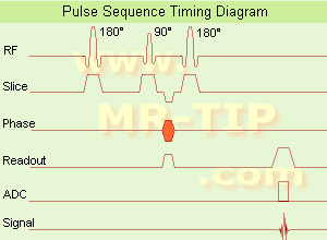
Inversion Recovery Sequence Timing Diagram (IR) The inversion recovery pulse sequence produces signals, which represent the longitudinal magnetization existing after the application of a 180° radio frequency pulse that rotates the magnetization Mz into the negative plane. After an inversion time (TI - time between the starting 180° pulse and the following 90° pulse), a further 90° RF pulse tilts some or all of the z-magnetization into the xy-plane, where the signal is usually rephased with a 180° pulse as in the spin echo sequence. During the initial time period, various tissues relax with their intrinsic T1 relaxation time.
In the pulse sequence timing diagram, the basic inversion recovery sequence is illustrated. The 180° inversion pulse is attached prior to the 90° excitation pulse of a spin echo acquisition. See also the Pulse Sequence Timing Diagram. There you will find a description of the components.
The inversion recovery sequence has the advantage, that it can provide very strong contrast between tissues having different T1 relaxation times or to suppress tissues like fluid or fat. But the disadvantage is, that the additional inversion radio frequency RF pulse makes this sequence less time efficient than the other pulse sequences.

Contrast values:
PD weighted: TE: 10-20 ms, TR: 2000 ms, TI: 1800 ms
T1 weighted: TE: 10-20 ms, TR: 2000 ms, TI: 400-800 ms
T2 weighted: TE: 70 ms, TR: 2000 ms, TI: 400-800 ms

See also Inversion Recovery, Short T1 Inversion Recovery, Fluid Attenuation Inversion Recovery, and Acronyms for 'Inversion Recovery Sequence' from different manufacturers.
 
Images, Movies, Sliders:
 Brain MRI Inversion Recovery  Open this link in a new window
    
 Knee MRI Sagittal STIR 002  Open this link in a new window
 Brain MRI Coronal FLAIR 001  Open this link in a new window
    
 
spacer

• View the DATABASE results for 'Inversion Recovery Sequence' (8).Open this link in a new window

 
Further Reading:
  Basics:
The equation for a repeated inversion recovery sequence
Contrast mechanisms in magnetic resonance imaging
2004   by www.iop.org    
  News & More:
FLAIR Vascular Hyperintensity: An Important MRI Marker in Patients with Transient Ischemic Attack
Thursday, 14 July 2022   by www.dovepress.com    
MRI Resources 
Shoulder MRI - MRI Accidents - MRI Reimbursement - Pacemaker - Mobile MRI - Process Analysis
 
previous      26 - 30 (of 90)     next
Result Pages : [1 2 3]  [4 5 6 7 8 9 10 11 12 13 14 15 16 17 18]
 Random Page
 
Share This Page
FacebookTwitterLinkedIn

MR-TIP    
Community   
User
Pass
Forgot your UserID/Password ?    


Personalized protocols (age, gender, body habitus, etc.) lead to :
more automated planning 
improved patient comfort 
better diagnostics 
optimized image quality 
nothing 

Look
      Ups


MR-TIP.com uses cookies! By browsing MR-TIP.com, you agree to our use of cookies.

Magnetic Resonance - Technology Information Portal
Member of SoftWays' Medical Imaging Group - MR-TIP • Radiology-TIP • Medical-Ultrasound-Imaging • 
Copyright © 2003 - 2024 SoftWays. All rights reserved. [ 24 November 2024]
Terms of Use | Privacy Policy | Advertising
 [last update: 2024-02-26 03:41:00]