 | Info
Sheets |
| | | | | | | | | | | | | | | | | | | | | | | | |
 | Out-
side |
| | | | |
|
| | | | | |  |
Result : Searchterm 'stir' found in 0 term [ ] and 49 definitions [ ] and 49 definitions [ ] ]
| 1 - 5 (of 49) nextResult Pages :  [1 2 3 4 5 6 7 8 9 10] [1 2 3 4 5 6 7 8 9 10] |  | |  | Searchterm 'stir' was also found in the following services: | | | | |
|  |  |
| |
|


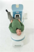
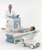
O-scan is manufactured and distributed by Esaote SpA
O-scan is a compact, dedicated extremity MRI system designed for easy installation and high throughput. The complete system fits in a 9' x 10' room, doesn't need for RF or magnetic shielding and it plugs in the wall. The 0.31T permanent magnet along with dual phased array RF coils, and advanced imaging protocols provide outstanding image quality and fast 25 minute complete examinations.
Esaote North America is the exclusive distributor of the O-scan system in the USA.
Device Information and Specification CLINICAL APPLICATION Dedicated Extremity
PULSE SEQUENCES
SE, HSE, HFE, GE, 2dGE, ME, IR, STIR, Stir T2, GE STIR, TSE, TME, FSE STIR, FSE ( T1, T2), X-Bone, Turbo 3DT1, 3D SHARC, 3D SST1, 3D SST2 2D: 2mm - 10 mm, 3D: 0.6 - 10 mm POWER REQUIREMENTS 100/110/200/220/230/240 | |  | | | | • Share the entry 'O-SCAN™':    | | | | |
|  | |  |  |  |
| |
|
Fat suppression is the process of utilizing specific MRI parameters to remove the deleterious effects of fat from the resulting images , e.g. with STIR, FAT SAT sequences, water selective (PROSET WATS - water only selection, also FATS - fat only selection possible) excitation techniques, or pulse sequences based on the Dixon method.
Spin magnetization can be modulated by using special RF pulses. CHESS or its variations like SPIR, SPAIR ( Spectral Selection Attenuated Inversion Recovery) and FAT SAT use frequency selective excitation pulses, which produce fat saturation.
Fat suppression techniques are nearly used in all body parts and belong to every standard MRI protocol of joints like knee, shoulder, hips, etc.

Image Guidance
Imaging of, e.g. the foot can induce bad fat suppression with SPIR/FAT SAT due to the asymmetric volume of this body part. The volume of the foot alters the magnetic field to a different degree than the smaller volume of the lower leg affecting the protons there. There is only a small band of tissue where the fat protons are precessing at the frequency expected, resulting in frequency selective fat saturation working only in that area. This can be corrected by volume shimming or creating a more symmetrical volume being imaged with water bags.
Even with their longer scan time and motion sensitivity, STIR (short T1/tau inversion recovery) sequences are often the better choice to suppress fat. STIR images are also preferred because of the decreased sensitivity to field inhomogeneities, permitting larger fields of views when compared to fat suppressed images and the ability to image away from the isocenter. See also Knee MRI.
Sequences based on Dixon turbo spin echo ( fast spin echo) can deliver a significant better fat suppression than conventional TSE/FSE imaging.
| | | |  | |
• View the DATABASE results for 'Fat Suppression' (28).
| | | | |  Further Reading: Further Reading: | | Basics:
|
|
News & More:
| |
| |
|  | |  |  |  |
| |
|
Device Information and Specification CLINICAL APPLICATION Whole body SE, FE, IR, STIR, FFE, DEFFE, DESE, TSE, DETSE, Single shot SE, DRIVE, Balanced FFE, MRCP, Fluid Attenuated Inversion Recovery, Turbo FLAIR, IR-TSE, T1- STIR TSE, T2- STIR TSE, Diffusion Imaging, 3D SE, 3D FFE, Contrast Perfusion Analysis, MTC;; Angiography: CE-ANGIO, MRA 2D, 3D TOFOpen x 47 cm x infinite (side-first patient entry) POWER REQUIREMENTS 400/480 V | |  | |
• View the DATABASE results for 'Panorama 0.6T™' (2).
| | | | |
|  |  | Searchterm 'stir' was also found in the following services: | | | | |
|  |  |
| |
|

From Philips Medical Systems;
this active shielded member of the Panorama product line combines the advantages of one 1.0 T system's with the possibilities of an open MRI system. The open design helps ease anxiety for claustrophobic patients and increased patient comfort whereby the field strength provides spectacular image quality and fast patient throughput.
Device Information and Specification CLINICAL APPLICATION Whole body Vertically opposed solenoids, head, head-neck, extremity, neck, body/ spine M-XL, shoulder, bilateral breast, wrist, TMJ, flex XS-S-M-L-XL-XXL SE, FE, IR, STIR, FFE, DEFFE, DESE, TSE, DETSE, Single shot SE, DRIVE, Balanced FFE, MRCP, FLAIR, Turbo FLAIR, IR-TSE, T1- STIR TSE, T2- STIR TSE, Diffusion Imaging, 3D SE, 3D FFE, Contrast Perfusion Analysis, MTC;; Angiography: CE-ANGIO, MRA 2D, 3D TOFOpen x 47 cm x infinite (side-first patient entry) POWER REQUIREMENTS 400/480 V | |  | |
• View the DATABASE results for 'Panorama 1.0T™' (2).
| | | | |
|  | |  |  |  |
| |
|
Quick Overview
DESCRIPTION
Signal loss, intensity variations

Image Guidance
| |  | | | |
|  | |  |  |
|  | |
|  | | |
|
| |
 | Look
Ups |
| |