 | Info
Sheets |
| | | | | | | | | | | | | | | | | | | | | | | | |
 | Out-
side |
| | | | |
|
| | | | | |  | Searchterm 't1' was also found in the following services: | | | | |
|  |  |
| |
|
Resovist® is an organ-specific MRI contrast agent, used for the detection and characterization of especially small focal liver lesions.
Resovist® consists of superparamagnetic iron oxide ( SPIO) nanoparticles coated with carboxydextran, which are accumulated by phagocytosis in cells of the reticuloendothelial system (RES) of the liver. The uptake of Resovist® Injection in the reticuloendothelial cells results in a decrease of the signal intensity of normal liver parenchyma on both T2- and T1 weighted images.
Most malignant liver tumors do not contain RES cells and therefore do not uptake the iron particles. The resulting imaging effect is an improved contrast between the tumor (bright) and the surrounding tissue (dark).
Resovist® can be injected as an intravenous bolus, which allows immediate imaging of the liver and reduces the overall examination time. A dynamic imaging strategy after bolus injection supports to characterize lesions.
In comprehensive clinical trials, it demonstrated an excellent safety profile.
In 2001, Resovist® was approved for the European market.
See also Superparamagnetic Iron Oxide.
Resovist® competed with Primovist™, the other liver imaging agent of Bayer Schering Pharma AG. Due to this reason, the production of Resovist® has been abandoned in 2009.
Drug Information and Specification T2/T1, Predominantly negative enhancement PHARMACOKINETIC RES-directed CONCENTRATION 0.5 mol Fe/L DOSAGE Less than 60 kg = 0.9 ml, greater than 60 kg = 1.4 ml PREPARATION Finished product PRESENTATION
Pre-filled syringes of 0.9 and 1.4 mL DO NOT RELY ON THE INFORMATION PROVIDED HERE, THEY ARE
NOT A SUBSTITUTE FOR THE ACCOMPANYING PACKAGE INSERT! Distribution Information TERRITORY TRADE NAME DEVELOPMENT
STAGE DISTRIBUTOR Australia Resovist® Approved - | |  | | | |  Further Reading: Further Reading: | News & More:
|
|
| |
|  | |  |  |  |
| |
|

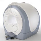
From GE Healthcare;
The Signa HDx MRI system is GE's leading edge whole body magnetic resonance scanner designed to support high resolution, high signal to noise ratio, and short scan times.
Signa HDx 3.0T offers new technologies like ultra-fast image reconstruction through the new XVRE recon engine, advancements in parallel imaging algorithms and the broadest range of premium applications. The HD applications, PROPELLER (high-quality brain imaging extremely resistant to motion artifacts), TRICKS (contrast-enhanced angiographic vascular lower leg imaging), VIBRANT (for breast MRI), LAVA (high resolution liver imaging with shorter breath holds and better organ coverage) and MR Echo (high-definition cardiac images in real time) offer unique capabilities.
Device Information and Specification CLINICAL APPLICATION Whole body
CONFIGURATION Compact short bore SE, IR, 2D/3D GRE, RF-spoiled GRE, 2DFGRE, 2DFSPGR, 3DFGRE, 3DFSPGR, 3DTOFGRE, 3DFSPGR, 2DFSE, 2DFSE-XL, 2DFSE-IR, T1-FLAIR, SSFSE, EPI, DW-EPI, BRAVO, Angiography: 2D/3D TOF, 2D/3D phase contrast vascular IMAGING MODES Single, multislice, volume study, fast scan, multi slab, cine, localizer H*W*D 240 x 2216,6 x 201,6 cm POWER REQUIREMENTS 480 or 380/415, 3 phase ||
COOLING SYSTEM TYPE Closed-loop water-cooled grad. | |  | | | |
|  | |  |  |  |
| |
|

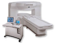
From GE Healthcare;
the New Signa Profile/i is a patient friendly open MRI system that virtually eliminates patient anxiety and claustrophobia, without compromising diagnostic utility.
Device Information and Specification CLINICAL APPLICATION Whole body Integrated transmit body coil, body flex sizes: M, L, XL, quadrature, head coil quadrature, 4 channel neurovascular array, 8 channel CTL array, quad. c- spine, 2 channel shoulder array, extremity coil, 3 channel wrist array, 4 channel breast array, 6, 9, 11 inch general purpose loop coils Standard: SE, IR, 2D/3D GRE and SPGR, Angiography: 2D/3D TOF, 2D/3D phase contrast; 2D/3D FSE, 2D/3D FRFSE, FGRE and FSPGR, SSFP, FLAIR, EPI, optional: 2D/3D Fiesta, fat/water separation, T1 FLAIRIMAGING MODES Localizer, single slice, multislice, volume, fast, POMP, multi slab, cine, slice and frequency zip, extended dynamic range, tailored RF TR 6 to 12000 msec in increments of 1 msec TE 1.3 to 2000 msec in increments of 1 msec 2D: 2.7mm - 20mm 3D: 0.2mm - 5mm 0.08 mm; 0.02 mm optional 10,000 kg w/gradient enclosure POWER REQUIREMENTS 200 - 480, 3-phase COOLING SYSTEM TYPE None required | |  | |
• View the DATABASE results for 'Signa Profile™' (2).
| | | | |
|  |  | Searchterm 't1' was also found in the following services: | | | | |
|  | |  | |  |  |  |
| |
|
A method used to trace the motion or flow of tissue. Nuclei will retain their magnetic orientation for a time on the order of T1 even in the presence of motion. Thus, if the nuclei in a given region have their spin orientation changed, the altered spins will serve as a 'tag' to trace the motion for a time on the order of T1 of any fluid that may have been in the tagged region. | |  | |
• View the DATABASE results for 'Spin Tagging' (3).
| | | | |
|  | |  |  |
|  | |
|  | | |
|
| |
 | Look
Ups |
| |