|
Magnetic
Resonance -
Technology
Information
Portal |
Welcome to MRI Technology • • |
|
|
 | Info
Sheets |
| | | | | | | | | | | | | | | | | | | | | | | | |
 | Out-
side |
| | | | |
|
| | | | | |
| Result: Searchterm 'AIN'
found in 209 messages |
| Result Pages: 1 2 3 4 5 6 7 8 9 10 11 [12] 13 14 15 16 17 18 19 20 21 22 23 24 25 26 27 28 29 30 31 32 33 34 35 36 37 38 39 40 41 42 |
More Results:  Database (389) News Service (888) Resources (128) Database (389) News Service (888) Resources (128) |
|
|
Bob Smtih
Thu. 30 Oct.14,
18:41
[Start of:
'What is this white "cloud" on the right side of this MRI?'
0 Reply]
 Category: Category:
General
|
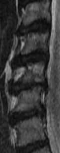
| What is this white "cloud" on the right side of this MRI? |
The following was noted:c2/c3 normal c3/c4 Very minimal anterior ostepphytic ridging is present. There is no spinal stenosis or neural foraminal narrowing. C4/c5 Very minimal anterior ostepphytic ridging is noted. There is no spinal stenosis or neural foraminal narrowing. c5/c6 There is a circumferential disc osteophyte complex present. There is mild central spinal stenosis. The AP diameter of the spinal canal is narrowed to 9mm. The dorsal and ventral CSF spaces remain patent. There is mild to moderate neural foraminal narrowing. c6/c7 a circumferential disc osteophyte complex present. There is mild central spinal stenosis. AP diameter of the spinal canal is narrowed to 9mm. The dorsal and ventral CSF spaces remain patent. There is moderate to severe neural foraminal narrowing. c7/t1 Normal. AP diameter of the spinal canal is 12 mm My question is C5 looks like it is chewed up; weird shading. Does it appear to be more damaged than the report states? Also there seems to be a white "cloud" that goes into the left side of multiple disks. What is the white "cloud"? c3 through c7 are in the image
 MRI SAG T2 MRI SAG T2
|
| |  Reply to this thread Reply to this thread
(login or register first) | |
Reader Mail
Wed. 29 Oct.14,
20:38
[Start of:
'Vibrant Sound Bridge and MRI'
0 Reply]
Category:
Devices, Scanner, Machines
|
| Vibrant Sound Bridge and MRI |
Hello everyone,
I have a Vibrant Soundbridge implanted on both sides of my skull, which contain a magnet. I use them due to severe hearing impairment.
Now the question is, I was told that MRI was prohibited for patients with a Vibrant Soundbridge.
Could anyone suggest an alternative to this problem please? Is there another equipment allowing me to have the conclusions normally obtained from MRI? My problem is an accident to my spinal cord, which requires further investigation using an MRI.
I would appreciate any help!
Thanks a lot
|
| | Reply to this thread
(login or register first) | |
michelle cyrus
Wed. 24 Sep.14,
16:43
[Start of:
'[URGENT] Need MO disk 2.3GB of Hitachi MRI AIRIS MATE'
0 Reply]
Category:
General
|
| [URGENT] Need MO disk 2.3GB of Hitachi MRI AIRIS MATE |
We are urgently looking for Maxell magneto optical disk 2.3GB 512byte/sector rewriteable MA172-S1.
The disk contains AIRIS MATE (ultraSPARC- IIi) system software.
Ship to: Vietnam
Please email us to singaporemedical@outlook.com or call +84912460068 if you have one.
Thanks a lot.
|
| | Reply to this thread
(login or register first) | |
Rena Miller
Fri. 15 Aug.14,
07:16
[Reply (1 of 2) to:
'door alarm/ MRI pauses if door opens'
started by: 'Samuel Strode'
on Thu. 7 Aug.14]
Category:
Devices, Scanner, Machines
|
| door alarm/ MRI pauses if door opens |
MRIs are enclosed by a Faraday cage to shield the MRI scanner against incoming radio waves which would disturb the sent and received signals. The reception or transmission of radio waves is heavily attenuated or blocked by the cage, which is usually integrated in the walls of the scanning room. The closed RF door completes the cage preventing RFI/EMI leakage, absorbs also much of the acoustic noise and prevents unauthorized persons to enter the room. Usually, all or the unreconstructed part of information is lost if the scan is stopped by door opening. Some MRI systems allow to pause the scan (at the keyboard, not by door opening) and to continue scanning while not loosing data.
|
| | View the whole thread | | |
Steven Ford
Tue. 5 Aug.14,
19:57
[Reply (3 of 4) to:
'MRI artifact'
started by: 'Daniel Cronk'
on Mon. 16 Jun.14]
Category:
Artifacts
|
| MRI artifact |
No it is not an RF leak artifact. That would appear different. I would guess that there was one instance of motion. Note that the artifact is mostly on the high contrast areas (white against black) where the patient can move the most.
Also it may be that your acquisition matrix was a little on the low side, which would lead to larger pixel-y appearance that is more apparent when there's motion.
Steven Ford
Professional Imaging Services, Inc.
|
| | View the whole thread | |
| |
| | Result Pages : 1 2 3 4 5 6 7 8 9 10 11 [12] 13 14 15 16 17 18 19 20 21 22 23 24 25 26 27 28 29 30 31 32 33 34 35 36 37 38 39 40 41 42 | |
|
| |
 | Look
Ups |
| |
|
MR-TIP.com uses cookies! By browsing MR-TIP.com, you agree to our use of cookies. | | | [last update: 2024-02-26 03:41:00] |
|
|