 |
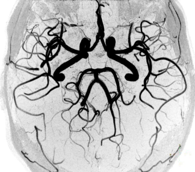
TOF-MRA Circle of Willis Inverted MIP |
|
 |
|
 |
 |
 | | This maximum intensity projection (MIP)in the coronal plane shows the brain vessel anatomy and blood supply. The internal carotid arteries (ICA - right and left thick vessels) and the vertebral arteries join together at the base of the brain. The connection of the vertebral arteries forms the basilar artery (seen in the lower, middle part of the picture).
| | | |  |
| |
| | |
|
 |
 |
 | | The MIP in transverse projection shows the circle of Willis, formed around the optic chiasm and the pituitary stalk (brain tissue is not seen due to MIP processing).Seen are the anterior communicating arteries (right and left ICA, right and left anterior cerebral arteries (ACA), anterior communicating artery (ACOM)) and the posterior communicating arteries (right and left posterior communicating arteries (PCOM), right and left posterior cerebral arteries (PCA), basilar artery). | | | |  |
| |
| | |
|
 |
| |
| | |
|
 |
| |
| | |
| |
| Related Images
• Brain MRI Inversion Recovery
• Brain MRI Coronal FLAIR 001
• Brain MRI Sagittal T1 001
• Brain MRI Transversal T1 001
• Brain MRI Transversal T1 002
• Brain MRI Transversal T2 001
• Brain MRI Transversal T2 002
• Brain MRI Transversal T2 003
• Brain MRI Images Axial T2
• Brain MRI Images T1
• PCA-MRA 3D Brain Venography Colored MIP |
 |
| Related Sliders
• MRI of the Skull Base
• MRI of the Brain Stem with Temoral Bone and Auditory System
• Circle of Willis, Time of Flight, MIP |
|
|
|
| |
| |
| Relevant Database Entries
• Angiography
• Brain MRI
• Circle of Willis
• Entry Slice Phenomenon (Artifact)
• Flow
• Flow Effects
• Flow Related Enhancement
• Inflow Magnetic Resonance Angiography
• Magnetic Resonance Angiography MRA
• Maximum Intensity Projection
• MRI
• Postprocessing
• Spin Phase Effect
• Time of Flight Angiography
• Vessel Separation |
| |