 | Info
Sheets |
| | | | | | | | | | | | | | | | | | | | | | | | |
 | Out-
side |
| | | | |
|
| | | | | |  |
|  |  |
 |
| |
|
 [This entry is marked for removal.]
E-Z-EM, Inc. is headquartered in New York and develops, manufactures and markets diagnostic imaging products used by radiologists like the MRI bowel marker Gadolite® Oral Suspension.
In addition to medical diagnostics systems, the company's products include MR-compatible interventional devices, such as biopsy needles. E-Z-EM is also a third-party contract manufacturer of contrast media.
Bracco Diagnostics, Inc. , the US-based subsidiary of Bracco Imaging S.p.A. and part of the Bracco Group, announced in Oct. 2007 that it has entered into a merger agreement to acquire E-Z-EM, Inc. (NASDAQ: EZEM) for a total consideration of about $ 240 million. Upon completion of the procedure and subject to the approval of the regulatory authorities, Bracco Diagnostics Inc. will incorporate E-Z-EM.
Contact Information
MAIL
E-Z-EM, Inc.
Westbury, New York
USA
| |  | |
• View the NEWS results for 'E-Z-EM, Inc.' (1).
| | | | | |  |
 |
| |
|
Device Information and Specification
CLINICAL APPLICATION
Whole body
SE, IR, FSE, FIR, GE, SG, BASG, PBSG, PCIR, DWI, Radial, Angiography: TOF, FLUTE (Fluoro-triggered bolus MRA), Time-resolved MRA
IMAGING MODES
Single, multislice, volume study
Level Range: -2,000 to +4,000
POWER REQUIREMENTS
208/220/240 V, single phase
| |  | |
• View the NEWS results for 'Echelon™ 1.5T' (3).
| | |
• View the DATABASE results for 'Echelon™ 1.5T' (2).
| | | | |  Further Reading: Further Reading: | Basics:
|
|
| | |  | |  |  |
 |
| |
| In MRI, an echo is the emission of energy in form of an electromagnetic resonance signal of a nuclei after its excitation. At this point spins are back in phase again and the signal is measured. The desired number of echoes is selectable. Often until eight echoes are permissible for 2D or 3D scans using spin echo, inversion recovery or MIX techniques. Two echoes are permissible for all other techniques. A multi echo imaging sequence is needed for simultaneous measurement of T2 and density weighted images. | |  | |
• View the NEWS results for 'Echo' (4).
| | |
• View the DATABASE results for 'Echo' (305).
| | | | |  Further Reading: Further Reading: | News & More:
|
|
| | |  |
 |
| |
| | |  | | | |  Further Reading: Further Reading: | News & More:
|
|
| | |  |
 |
| |
|

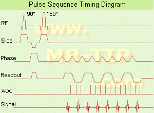
(EPI) Echo planar imaging is one of the early magnetic resonance imaging sequences (also known as Intascan), used in applications like diffusion, perfusion, and functional magnetic resonance imaging. Other sequences acquire one k-space line at each phase encoding step. When the echo planar imaging acquisition strategy is used, the complete image is formed from a single data sample (all k-space lines are measured in one repetition time) of a gradient echo or spin echo sequence (see single shot technique) with an acquisition time of about 20 to 100 ms.
The pulse sequence timing diagram illustrates an echo planar imaging sequence from spin echo type with eight echo train pulses. (See also Pulse Sequence Timing Diagram, for a description of the components.)
In case of a gradient echo based EPI sequence the initial part is very similar to a standard gradient echo sequence. By periodically fast reversing the readout or frequency encoding gradient, a train of echoes is generated.
EPI requires higher performance from the MRI scanner like much larger gradient amplitudes. The scan time is dependent on the spatial resolution required, the strength of the applied gradient fields and the time the machine needs to ramp the gradients.
In EPI, there is water fat shift in the phase encoding direction due to phase accumulations. To minimize water fat shift (WFS) in the phase direction fat suppression and a wide bandwidth (BW) are selected. On a typical EPI sequence, there is virtually no time at all for the flat top of the gradient waveform. The problem is solved by "ramp sampling" through most of the rise and fall time to improve image resolution.
The benefits of the fast imaging time are not without cost. EPI is relatively demanding on the scanner hardware, in particular on gradient strengths, gradient switching times, and receiver bandwidth. In addition, EPI is extremely sensitive to image artifacts and distortions. | |  | |
• View the NEWS results for 'Echo Planar Imaging' (1).
| | |
• View the DATABASE results for 'Echo Planar Imaging' (19).
| | | | |  Further Reading: Further Reading: | Basics:
|
|
| | |  | |  |  |
| | |
|
| |
 | Look
Ups |
| |