Magnetic Resonance - Technology Information Portal Welcome to MRI Technology
Info
  Sheets

Out-
      side
 

MRI is trending to low field magnets :
reduced costs will lead to this change 
AI will close the gap to high field 
only in remote areas 
is only temporary 
never 


MRI Sequences


ball_redSpin Echo Sequence 
Spin Echo Timing Diagram (SE) The most common pulse sequence used in MR imaging is based of the detection of a spin or Hahn echo. It uses 90° radio frequency pulses to excite the magnetization and one or more 180° pulses to refocus the spins to generate signal echoes named spin echoes (SE).
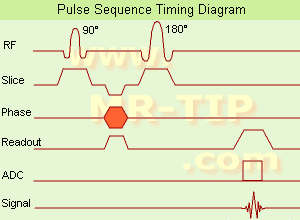
In the pulse sequence timing diagram, the simplest form of a spin echo sequence is illustrated.
The 90° excitation pulse rotates the longitudinal magnetization (Mz) into the xy-plane and the dephasing of the transverse magnetization (Mxy) starts.
The following application of a 180° refocusing pulse (rotates the magnetization in the x-plane) generates signal echoes. The purpose of the 180° pulse is to rephase the spins, causing them to regain coherence and thereby to recover transverse magnetization, producing a spin echo.
The recovery of the z-magnetization occurs with the T1 relaxation time and typically at a much slower rate than the T2-decay, because in general T1 is greater than T2 for living tissues and is in the range of 100-2000 ms.
The SE pulse sequence was devised in the early days of NMR days by Carr and Purcell and exists now in many forms: the multi echo pulse sequence using single or multislice acquisition, the fast spin echo (FSE/TSE) pulse sequence, echo planar imaging (EPI) pulse sequence and the gradient and spin echo (GRASE) pulse sequence;; all are basically spin echo sequences.
In the simplest form of SE imaging, the pulse sequence has to be repeated as many times as the image has lines.
Contrast values:
PD weighted: Short TE (20 ms) and long TR.
T1 weighted: Short TE (10-20 ms) and short TR (300-600 ms)
T2 weighted: Long TE (greater than 60 ms) and long TR (greater than 1600 ms)
With spin echo imaging no T2* occurs, caused by the 180° refocusing pulse. For this reason, spin echo sequences are more robust against e.g., susceptibility artifacts than gradient echo sequences.

See also Pulse Sequence Timing Diagram to find a description of the components.

• View the DATABASE results for 'Spin Echo Sequence' (24).Open this link in a new window

 
Further Reading:
  Basics:
Fast Spin Echo(.pdf)
Tuesday, 24 January 2006   by www.81bones.net    
Magnetic resonance imaging
   by www.scholarpedia.org    
FUNDAMENTALS OF MRI: Part I
   by www.e-radiography.net    
  News & More:
New MR sequence helps radiologists more accurately evaluate abnormalities of the uterus and ovaries
Thursday, 23 April 2009   by www.eurekalert.org    
MRI techniques improve pulmonary embolism detection
Monday, 19 March 2012   by medicalxpress.com    
Dual Echo Sequence 
(DE - dual / double echo) Dual echo sequences include images with different weightings and / or echo times and are used to obtain both, proton density and T2 weighted images or in phase and out of phase gradient echo images, simultaneously without increasing the measurement time.

• View the DATABASE results for 'Dual Echo Sequence' (3).Open this link in a new window

 
Further Reading:
  Basics:
BASIC PRINCIPLES OF MR IMAGING
   by spinwarp.ucsd.edu    
MRI evaluation of fatty liver in day to day practice: Quantitative and qualitative methods
Wednesday, 3 September 2014   by www.sciencedirect.com    
  News & More:
Iron overload: accuracy of in-phase and out-of-phase MRI as a quick method to evaluate liver iron load in haematological malignancies and chronic liver disease
Friday, 1 June 2012   by www.ncbi.nlm.nih.gov    
Modified Spin Echo 
(MSE) A spin echo technique with a flip angle over 90°.

See Spin Echo Sequence and Fast Spin Echo.
Multi Echo Multiplanar 
(MEMP) Sequence with a multislice and multi echo acquisition in one TR.

See also Multi Echo Imaging, Multiple Echo Imaging and Fast Spin Echo.

• View the DATABASE results for 'Multi Echo Multiplanar' (2).Open this link in a new window

Partial Saturation Spin Echo 
(PSSE) Partial saturation sequence in which the signal is detected as a spin echo. Even though a spin echo is used, there will not necessarily be a significant contribution of the T2 relaxation time to image contrast, unless the echo time, TE, is on the order of or longer than T2.
Variable Echo Multiplanar 
(VEMP) MR imaging spin echo pulse sequence in which signals for multiple variable echoes are collected.

• View the DATABASE results for 'Variable Echo Multiplanar' (2).Open this link in a new window

  Fast Spin Echo top
As a general rule the most successful man in life is the man who has the best information.
- Benjamin Disraeli
 
Share This Page
FacebookTwitterLinkedIn

MR-TIP    
Community   
User
Pass
Forgot your UserID/Password ?    


Look
      Ups


MR-TIP.com uses cookies! By browsing MR-TIP.com, you agree to our use of cookies.

Magnetic Resonance - Technology Information Portal
Member of SoftWays' Medical Imaging Group - MR-TIP • Radiology-TIP • Medical-Ultrasound-Imaging • 
Copyright © 2003 - 2024 SoftWays. All rights reserved. [ 21 November 2024]
Terms of Use | Privacy Policy | Advertising
 [last update: 2024-02-26 03:41:00]