 | Info
Sheets |
| | | | | | | | | | | | | | | | | | | | | | | | |
 | Out-
side |
| | | | |
|
| | | | |
Result : Searchterm 'Array Coil' found in 4 terms [ ] and 18 definitions [ ] and 18 definitions [ ] ]
| | previous 16 - 20 (of 22) nextResult Pages : [1] [2 3 4 5] |  | |  | Searchterm 'Array Coil' was also found in the following services: | | | | |
| |  |
| |
|
MRI of the lumbar spine, with its multiplanar 3 dimensional imaging capability, is currently the preferred modality for establishing a diagnosis. MRI scans and magnetic resonance myelography have many advantages compared with computed tomography and/or X-ray myelography in evaluating the lumbar spine. MR imaging scans large areas of the spine without ionizing radiation, is noninvasive, not affected by bone artifacts, provides vascular imaging capability, and makes use of safer contrast agents ( gadolinium chelate).
Due to the high level of tissue contrast resolution, nerves and discs are clearly visible. MRI is excellent for detecting degenerative disease in the spine. Lumbar spine MRI accurately shows disc disease (prolapsed disc or slipped disc), the level at which disc disease occurs, and if a disc is compressing spinal nerves. Lumbar spine MRI depicts soft tissues, including the cauda equina, spinal cord, ligaments, epidural fat, subarachnoid space, and intervertebral discs. Loss of epidural fat on T1 weighted images, loss of cerebrospinal fluid signal around the dural sac on T2 weighted images and degenerative disc disease are common features of lumbar stenosis.
Common indications for MRI of the lumbar spine:
•
Neurologic deficits, evidence of radiculopathy, acute spinal cord compression (e.g., sudden bowel/bladder disturbance)
•
Suspected systemic disorders (primary tumors, drop metastases, osteomyelitis)
•
Postoperative evaluation of lumbar spine: disk vs. scar
•
Localized back pain with no radiculopathy (leg pain)
Lumbar spine imaging requires a special spine coil. often used whole spine array coils have the advantage that patients do not need other positioning if also upper parts of the spine should be scanned. Sagittal T1 and T2 weighted FSE sequences are the standard views. With multi angle oblique techniques individually oriented transverse images of each intervertebral disc at different angles can be obtained.
See also the related poll result: ' MRI will have replaced 50% of x-ray exams by' | | | | | | | | | | | | |  Further Reading: Further Reading: | | Basics:
|
|
News & More:
| |
| |
| | | | | |
| |
|
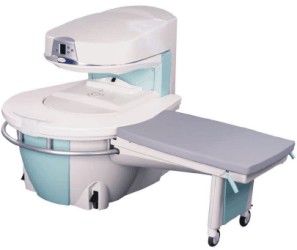
Manufactured by Esaote S.p.A.;
a low field open MRI scanner with permanent magnet for orthopedic use. The outstanding feature of this MRI system is a patient friendly design with 24 cm diameter, which allows the imaging of extremities and small body parts like shoulder MRI. The power consumption is around 1.3 kW and the needed minimum floor space is an area of 16 sq m.
At RSNA 2006 Hologic Inc. introduced a new dedicated extremity MRI scanner, the Opera. Manufactured by Esaote is the Opera a redesign of Esaote's 0.2 Tesla E-Scan XQ platform, which now enables complete imaging of all extremities, including hip and shoulder applications. 'Real-time positioning' reportedly speeds patient setup and reduces exam times.
Esaote North America and Hologic Inc are the U.S. distributors of this MRI device.
Device Information and Specification CLINICAL APPLICATION Dedicated extremity
SE, GE, IR, STIR, FSE, 3D CE, GE-STIR, 3D GE, ME, TME, HSE IMAGING MODES Single, multislice, volume study, fast scan, multi slab2D: 2 mm - 10 mm;
3D: 0.6 mm - 10 mm 4096 gray lvls, 256 lvls in 3D POWER REQUIREMENTS 2,0 kW; 110/220 V single phase | | | |
• View the DATABASE results for 'Opera (E-SCAN™ XQ)' (2).
| | | | | | Further Reading: | News & More:
|
|
| |
| | | | | |
| |
|
In parallel MR imaging, a reduced data set in the phase encoding direction(s) of k-space is acquired to shorten acquisition time, combining the signal of several coil arrays. The spatial information related to the phased array coil elements is utilized for reducing the amount of conventional Fourier encoding.
First, low-resolution, fully Fourier-encoded reference images are required for sensitivity assessment. Parallel imaging reconstruction in the Cartesian case is efficiently performed by creating one aliased image for each array element using discrete Fourier transformation. The next step then is to create an full FOV image from the set of intermediate images.
Parallel reconstruction techniques can be used to improve the image quality with increased signal to noise ratio, spatial resolution, reduced artifacts, and the temporal resolution in dynamic MRI scans.
Parallel imaging algorithms can be divided into 2 main groups:
Image reconstruction produced by each coil ( reconstruction in the image domain, after Fourier transform): SENSE ( Sensitivity Encoding), PILS (Partially Parallel Imaging with Localized Sensitivity),
ASSET.
Reconstruction of the Fourier plane of images from the frequency signals of each coil ( reconstruction in the frequency domain, before Fourier transform): GRAPPA. Additional techniques include SMASH, SPEEDER™,
IPAT (Integrated Parallel Acquisition Techniques - derived of GRAPPA a k-space based technique) and mSENSE (an image based enhanced version of SENSE).
| | | | | |
• View the DATABASE results for 'Parallel Imaging Technique' (12).
| | | | | | Further Reading: | Basics:
|
|
News & More:
| |
| |
| | | Searchterm 'Array Coil' was also found in the following services: | | | | |
| | |
| |
|
A coil is a large inductor with a considerable dimension and a defined wavelength, commonly used in configurations for MR imaging. The frequency of the radio frequency coil is defined by the Larmor relationship. The MRI image quality depends on the signal to noise ratio (SNR) of the acquired signal from the patient. Several MR imaging coils are necessary to handle the diversity of applications. Large coils have a large measurement field, but low signal intensity and vice versa (see also coil diameter). The closer the coil to the object, the stronger the signal - the smaller the volume, the higher the SNR. SNR is very important in obtaining clear images of the human body. The shape of the coil depends on the image sampling. The best available homogeneity can be reached by choice of the appropriate coil type and correct coil positioning. Orientation is critical to the sensitivity of the RF coil and therefore the coil should be perpendicular to the static magnetic field.
RF coils can be differentiated by there function into three general categories:
The RF signal is in the range of 10 to 100 MHz. During a typical set of clinical image measurements, the entire frequency spectrum of interest is of the order 10 kHz, which is an extremely narrow band, considering that the center frequency is about 100 MHz. This allows the use of single-frequency matching techniques for coils because their inherent bandwidth always exceeds the image bandwidth. The multi turn solenoid, bird cage coil, single turn solenoid, and saddle coil are typically operated as the transmitter and receiver of RF energy. The surface and phased array coils are typically operated as a receive only coil.
See also the related poll result: ' 3rd party coils are better than the original manufacturer coils' | | | | | |
• View the DATABASE results for 'Radio Frequency Coil' (9).
| | | | | | Further Reading: | Basics:
|
|
News & More:
| |
| |
| | | | | |
| |
|
| | | |
• View the DATABASE results for 'Sense Coil' (2).
| | | | | | Further Reading: | Basics:
|
|
| |
| | | | |
| | | |
|
| |
 | Look
Ups |
| |


