 | Info
Sheets |
| | | | | | | | | | | | | | | | | | | | | | | | |
 | Out-
side |
| | | | |
|
| | | 'Automatic Bolus Detection' | |
Result : Searchterm 'Automatic Bolus Detection' found in 1 term [ ] and 4 definitions [ ] and 4 definitions [ ] ]
| 1 - 5 (of 5) Result Pages :  [1] [1] |  | | |  |  |  |
| Automatic Bolus Detection |  |
| |
|
| |  | | | | • Share the entry 'Automatic Bolus Detection':    | | | | |  Further Reading: Further Reading: | | Basics:
|
|
News & More:
| |
| |
|  | |  |  |  |
| |
|
(CE MRA) Contrast enhanced MR angiography is based on the T1 values of blood, the surrounding tissue, and paramagnetic contrast agent.
T1-shortening contrast agents reduces the T1 value of the blood (approximately to 50 msec, shorter than that of the surrounding tissues) and allow the visualization of blood vessels, as the images are no longer dependent primarily on the inflow effect of the blood.
Contrast enhanced MRA is performed with a short TR to have low signal (due to the longer T1) from the stationary tissue, short scan time to facilitate breath hold imaging, short TE to minimize T2* effects and a bolus injection of a sufficient dose of a gadolinium chelate.
Images of the region of interest are performed with 3D spoiled gradient echo pulse sequences. The enhancement is maximized by timing the contrast agent injection such that the period of maximum arterial concentration corresponds to the k-space acquisition. Different techniques are used to ensure optimal contrast of the arteries e.g., bolus timing, automatic bolus detection, bolus tracking, care bolus.
A high resolution with near isotropic voxels and minimal pulsatility and misregistration artifacts should be striven for. The postprocessing with the maximum intensity projection ( MIP) enables different views of the 3D data set.
Unlike conventional MRA techniques based on velocity dependent inflow or phase shift techniques, contrast enhanced MRA exploits the
gadolinium induced T1-shortening effects. CE MRA reduces or eliminates most of the artifacts of time of flight angiography or phase contrast angiography. Advantages are the possibility of in plane imaging of the blood vessels, which allows to examine large parts in a short time and high resolution scans in one breath hold.
CE MRA has found a wide acceptance in the clinical routine, caused by the
advantages:
•
3D MRA can be acquired in any plane, which means that
greater vessel coverage can be obtained at high
resolution with fewer slices (aorta, peripheral vessels);
•
the possibility to perform a time resolved examination
(similarly to conventional angiography);
•
no use of ionizing radiation; paramagnetic agents have a beneficial safety.
| | | |  | |
• View the DATABASE results for 'Contrast Enhanced Magnetic Resonance Angiography' (14).
| | |
• View the NEWS results for 'Contrast Enhanced Magnetic Resonance Angiography' (2).
| | | | |  Further Reading: Further Reading: | Basics:
|
|
News & More:
| |
| |
|  | |  |  |  |
| |
|
| |  | |
• View the DATABASE results for 'Fluoroscopic Triggering' (2).
| | | | |  Further Reading: Further Reading: | Basics:
|
|
| |
|  | |  |  |  |
| |
|

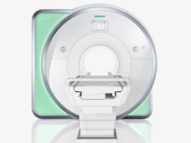
From Siemens Medical Systems;
Received FDA clearance in 2010.
The MAGNETOM Aera is a patient friendly, comfortable 1.5 Tesla MRI system with advanced radio frequency chain.
The system is equipped with the Tim 4G and Dot system (Total imaging matrix + Day optimizing throughput), to enhance both productivity and image quality.
Tim 4G technology provides improved SNR. The standard system configuration of 48 radio frequency channels and 204 coil elements creates an imaging matrix that allows maximum use of coil elements at full field of view. Dot provides improved image consistency through new features like auto align, auto FoV and automatic bolus detection.
Device Information and Specification
CLINICAL APPLICATION
Whole body
Head, spine, torso/ body coil, neurovascular, cardiac, neck, shoulder, knee, wrist, foot//ankle and multi-purpose flex coils. Peripheral vascular, breast, shoulder. Up to 60% more SNR with Tim 4G.
CHANNELS (min. / max. configuration)
48, 64
MINIMUM TE
3-D GRE: 0.22 (256 matrix), Ultra-short TE
At isocenter: L-R 70 cm, A-P (with table) 55 cm
MAGNET WEIGHT (gantry included)
3121 kg
DIMENSION H*W*D (gantry included)
145 x 231 x 219 cm
MAX. AMPLITUDE
33 or 45 mT/m
3 linear with 20 coils, 5 nonlinear 2nd-order
POWER REQUIREMENTS
380 / 400 / 420 / 440 / 460 / 480 V, 3-phase + ground; 85 kVA
| |  | | | |
|  | |  |  |  |
| |
|
| |  | |
• View the DATABASE results for 'Time Resolved Imaging of Contrast Kinetics' (2).
| | | | |  Further Reading: Further Reading: | Basics:
|
|
| |
|  | |  |  |
|  | 1 - 5 (of 5) Result Pages :  [1] [1] |
| |
|
| |
 | Look
Ups |
| |