Magnetic Resonance - Technology Information Portal Welcome to MRI Technology
Info
  Sheets

Out-
      side


 'Cardiovascular Imaging' 
SEARCH FOR    
 
  2 3 5 A B C D E F G H I J K L M N O P Q R S T U V W X Y Z
Result : Searchterm 'Cardiovascular Imaging' found in 1 term [] and 17 definitions [], (+ 7 Boolean[] results
previous     11 - 15 (of 25)     next
Result Pages : [1]  [2 3 4]  [5]
Searchterm 'Cardiovascular Imaging' was also found in the following services: 
spacer
News  (14)  Resources  (16)  
 
Echelonâ„¢ 1.5TInfoSheet: - Devices -
Intro, 
Types of Magnets, 
Overview, 
etc.
 
www.hitachimed.com/contentindex.asp?ID=971 From Hitachi Medical Systems America Inc.;
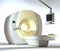
Hitachi expanded its portfolio with the Echelon™ 1.5T. The MRI scanner combines a compact magnet and a scalable 8-channel RF system with high-performance gradients and slew rate to select short echo times, small field of views, high matrices and thin slices. Standard features of the Echelon MRI system include higher-order active shim, RAPID (parallel imaging for use on brain MRI, body, cardiovascular imaging, and orthopedic coils), multiple coil ports, and an advanced reconstruction engine.
Device Information and Specification
CLINICAL APPLICATION
Whole body
CONFIGURATION
Short bore
Head, body coil, spine, breast, knee, shoulder, vascular multiple array coils.
SYNCHRONIZATION
Cardiac gating, ECG/peripheral, respiratory gating
PULSE SEQUENCES
SE, IR, FSE, FIR, GE, SG, BASG, PBSG, PCIR, DWI, Radial, Angiography: TOF, FLUTE (Fluoro-triggered bolus MRA), Time-resolved MRA
IMAGING MODES
Single, multislice, volume study
PIXEL INTENSITY
Level Range: -2,000 to +4,000
Sub millimeter
POWER REQUIREMENTS
208/220/240 V, single phase
CRYOGEN USE
Low cryogen boil-off
STRENGTH
30 mT/m
150 T/m/sec
Higher-order active shim
spacer
 
• Related Searches:
    • MRI Equipment
    • Superconducting Magnet
    • Excelart AGâ„¢ with Pianissimo
    • Hardware
    • MRI
 
Further Reading:
  Basics:
Echelon 1.5T
   by www.hitachimed.com    
Searchterm 'Cardiovascular Imaging' was also found in the following service: 
spacer
Radiology  (4) Open this link in a new window
FlowForum -
related threads
 
Flow phenomena are intrinsic processes in the human body. Organs like the heart, the brain or the kidneys need large amounts of blood and the blood flow varies depending on their degree of activity. Magnetic resonance imaging has a high sensitivity to flow and offers accurate, reproducible, and noninvasive methods for the quantification of flow. MRI flow measurements yield information of blood supply of of various vessels and tissues as well as cerebro spinal fluid movement.
Flow can be measured and visualized with different pulse sequences (e.g. phase contrast sequence, cine sequence, time of flight angiography) or contrast enhanced MRI methods (e.g. perfusion imaging, arterial spin labeling).
The blood volume per time (flow) is measured in: cm3/s or ml/min. The blood flow-velocity decreases gradually dependent on the vessel diameter, from approximately 50 cm per second in arteries with a diameter of around 6 mm like the carotids, to 0.3 cm per second in the small arterioles.

Different flow types in human body:
•
Behaves like stationary tissue, the signal intensity depends on T1, T2 and PD = Stagnant flow
•
Flow with consistent velocities across a vessel = Laminar flow
•
Laminar flow passes through a stricture or stenosis (in the center fast flow, near the walls the flow spirals) = Vortex flow
•
Flow at different velocities that fluctuates = Turbulent flow

See also Flow Effects, Flow Artifact, Flow Quantification, Flow Related Enhancement, Flow Encoding, Flow Void, Cerebro Spinal Fluid Pulsation Artifact, Cardiovascular Imaging and Cardiac MRI.
 
Images, Movies, Sliders:
 MVP Parasternal  Open this link in a new window
    

Courtesy of  Robert R. Edelman
 TOF-MRA Circle of Willis Inverted MIP  Open this link in a new window
    

 Circle of Willis, Time of Flight, MIP  Open this link in a new window
    
SlidersSliders Overview

 
spacer

• View the DATABASE results for 'Flow' (113).Open this link in a new window


• View the NEWS results for 'Flow' (7).Open this link in a new window.
 
Further Reading:
  News & More:
The super-fast MRI scan that could revolutionise heart failure diagnosis
Wednesday, 21 September 2022   by www.eurekalert.org    
MRI Resources 
Safety pool - Safety Products - Intraoperative MRI - Shielding - Universities - Pacemaker
 
Heart MRI
 
Magnetic resonance imaging of the heart is a diagnostic MRI procedure that is useful to evaluate the structures, the function and viability of the heart and the major blood vessels.

See also Cardiac MRI, Cardiovascular Imaging, MRA, Cardiac Stent, Myocardial Late Enhancement and Coronary Angiography.
Radiology-tip.comradHeart Scintigraphy,  Coronary CT Angiography
spacer
Medical-Ultrasound-Imaging.comCardiac Ultrasound,  Echocardiography
spacer

• View the NEWS results for 'Heart MRI' (18).Open this link in a new window.
 
Further Reading:
  Basics:
Advanced Imaging Can ID More Causes Of Stroke Before They Strike
Thursday, 22 March 2007   by www.sciencedaily.com    
New technique could allow for safer, more accurate heart scans
Thursday, 10 December 2015   by www.gizmag.com    
  News & More:
Cardiac MRI of Hereditary Cardiomyopathy
Friday, 11 March 2022   by pubs.rsna.org    
MRI sheds light on COVID vaccine-associated heart muscle injury
Tuesday, 15 February 2022   by www.sciencedaily.com    
Manganese enhanced MRI provides more accurate details of heart function after a heart attack
Tuesday, 11 May 2021   by www.news-medical.net    
Cardiac MRI Becoming More Widely Available Thanks to AI and Reduced Exam Times
Wednesday, 19 February 2020   by www.dicardiology.com    
Tiny Brain Blocks as Marker for Heart Disease Too?
Monday, 6 February 2017   by www.medpagetoday.com    
MRI shows heart ages differently in women than in men
Tuesday, 20 October 2015   by www.eurekalert.org    
Brains Of Term Infants With Heart Disease Resemble Those Of Preemies
Monday, 12 November 2007   by www.sciencedaily.com:80    
Searchterm 'Cardiovascular Imaging' was also found in the following services: 
spacer
News  (14)  Resources  (16)  
 
Intera 3.0TPanorama 0.2InfoSheet: - Devices -
Intro, 
Types of Magnets, 
Overview, 
etc.MRI Resource Directory:
 - Devices -
 
www.medical.philips.com/de/products/mri/products/ From Philips Medical Systems;
the Intera 3 T high field system, the first with a compact magnet, which is built on the same platform as the 1.5 T, is targeted to high-end neurological, orthopedic and cardiovascular imaging applications with maximum patient comfort and acceptance without compromising image quality and clinical performance. Useable for clinical routine and research. The Intera systems offer diffusion tensor imaging (DTI) fiber tracking that measures movement of water in the brain and can therefore detect areas of the brain where normal movement of water is disrupted.
Device Information and Specification
CLINICAL APPLICATION
Whole body
CONFIGURATION
Short bore compact
Standard: head, body, C1, C3; Optional: Small joint, flex-E, flex-R, endocavitary (L and S), dual TMJ, knee, neck, T/L spine, breast; Optional phased array: spine;; Optional SENSE coils: Flex body, flex cardiac, neuro-vascular, head
Optional
SYNCHRONIZATION
ECG/peripheral: Optional/yes, respiratory gating
PULSE SEQUENCES
SE, Modified-SE, IR (T1, T2, PD), STIR, FLAIR, SPIR, FFE, T1-FFE, T2-FFE, Balanced FFE, TFE, Balanced TFE, Dynamic, Keyhole, 3D, Multi Chunk 3D, Multi Stack 3D, K Space Shutter, MTC, TSE, Dual IR, DRIVE, EPI, Cine, 2DMSS, DAVE, Mixed Mode; Angiography: Inflow MRA, TONE, PCA, CE MRA
IMAGING MODES
Single Slice 2D , Multi Single Slice 2D, Multi Slice 2D, 3D, Multi Chunk 3D, Multi Stack 3D
TR
Min. 1.6 (Master) msec
TE
Min. 0.5 (Master) msec
SINGLE/MULTI SLICE
RapidView Recon. greater than 500 @ 256 Matrix
FOV
Max. 53 cm
0.1 mm (Omni), 0.05 mm (Power)
128 x 128, 256 x 256,512 x 512,1024 x 1024 (64 for Bold img)
MEASURING MATRIX
Variable in 1% increments
PIXEL INTENSITY
Lum.: 120 cd/m2; contrast: 150:1
Variable (op. param. depend.)
60 x 60 cm
MAGNET WEIGHT
5500 kg
H*W*D
240 x 188 x 157 cm
POWER REQUIREMENTS
380/400 V
CRYOGEN USE
0.07 L/hr helium
STRENGTH
30 (Master) mT/m
5-GAUSS FRINGE FIELD
3.0 m / 5.2 m
Passive and dynamic
spacer

• View the DATABASE results for 'Intera 3.0T™' (2).Open this link in a new window

Searchterm 'Cardiovascular Imaging' was also found in the following service: 
spacer
Radiology  (4) Open this link in a new window
Intera Achieva CVâ„¢InfoSheet: - Devices -
Intro, 
Types of Magnets, 
Overview, 
etc.MRI Resource Directory:
 - Devices -
 
www.medical.philips.com/main/products/mri/products/intera_achievacv/ From Philips Medical Systems;
The Intera Achieva CV is focused on cardiovascular imaging research and development, generating technology on the cutting edge for modern clinical needs, like easy coronary artery imaging, the evaluation of flow effects in vessels and peripheral angiography with optimal resolution per station.
Device Information and Specification
CLINICAL APPLICATION
Whole body
CONFIGURATION
Short bore compact
Standard: Head, body, cardiac, optional phased array: Spine, pediatric, 3rd party connector; Optional SENSE? coils for all applications
Optional
SYNCHRONIZATION
ECG/peripheral: Optional/yes, respiratory gating
PULSE SEQUENCES
SE, Modified-SE, IR (T1, T2, PD), STIR, FLAIR, SPIR, FFE, T1-FFE, T2-FFE, Balanced FFE, TFE, Balanced TFE, Dynamic, Keyhole, 3D, Multi Chunk 3D, Multi Stack 3D, K Space Shutter, MTC, TSE, Dual IR, DRIVE, EPI, Cine, 2DMSS, DAVE, Mixed Mode; Angiography: Inflow MRA, TONE, PCA, CE MRA
IMAGING MODES
Single Slice 2D , Multi Single Slice 2D, Multi Slice 2D, 3D, Multi Chunk 3D, Multi Stack 3D
FOV
Over 50 cm
0.05 mm
128 x 128, 256 x 256,512 x 512,1024 x 1024 (64 for Bold img)
MEASURING MATRIX
Variable in 1% increments
PIXEL INTENSITY
Lum.: 120 cd/m2; contrast: 150:1
Variable (op. param. depend.)
60 x 60 cm
POWER REQUIREMENTS
380/400 V
CRYOGEN USE
0.03 L/hr helium
STRENGTH
up to 33 mT/m
5-GAUSS FRINGE FIELD
2.4 m / 3.8 m
Passive and dynamic
spacer

• View the DATABASE results for 'Intera Achieva CV™' (2).Open this link in a new window

 
Further Reading:
  News & More:
Coronary Artery Disease: Combined Stress MR Imaging Protocol-One-Stop Evaluation of Myocardial Perfusion and Function1
   by radiology.rsnajnls.org    
MRI Resources 
MRI Reimbursement - Stent - Supplies - Spectroscopy pool - Absorption and Emission - MR Myelography
 
previous      11 - 15 (of 25)     next
Result Pages : [1]  [2 3 4]  [5]
 Random Page
 
Share This Page
FacebookTwitterLinkedIn

MR-TIP    
Community   
User
Pass
Forgot your UserID/Password ?    


Next big thing in MRI will be :
AI 
remote operator 
personalized protocols 
helium-free 
molecular MRI 
portable MRI 

Look
      Ups


MR-TIP.com uses cookies! By browsing MR-TIP.com, you agree to our use of cookies.

Magnetic Resonance - Technology Information Portal
Member of SoftWays' Medical Imaging Group - MR-TIP • Radiology-TIP • Medical-Ultrasound-Imaging • 
Copyright © 2003 - 2024 SoftWays. All rights reserved. [ 21 November 2024]
Terms of Use | Privacy Policy | Advertising
 [last update: 2024-02-26 03:41:00]