 | Info
Sheets |
| | | | | | | | | | | | | | | | | | | | | | | | |
 | Out-
side |
| | | | |
|
| | | | | |  | Searchterm 'Cation' was also found in the following services: | | | | |
|  |  |
| |
|
Special imaging primarily means advanced MRI techniques used for qualitative and quantitative measurement of biological metabolism as e.g., spectroscopy, perfusion imaging (PWI, ASL), diffusion weighted imaging ( DWI, DTI, DTT) and brain function ( BOLD, fMRI). This physiological magnetic resonance techniques offer insights into brain structure, function, and metabolism.
Spectroscopy provides functional information related to identifi cation and quantifi cation of e.g. brain metabolites.
MR perfusion imaging has appli cations in stroke, trauma, and brain neoplasm. MRI provides the high spatial and temporal resolution needed to measure blood flow to the brain. arterial spin labeling techniques utilize the intrinsic protons of blood and brain tissue, labeled by special preparation pulses, rather than exogenous tracers injected into the blood.
MR diffusion tensor imaging characterizes the ability of water to spread across the brain in different directions. Diffusion parallel to nerve fibers has been shown to be greater than diffusion in the perpendicular direction. This provides a tool to study in vivo fiber connectivity in brain MRI.
FMRI allows the detection of a functional activation in the brain because cortical activity is intimately related to local metabolism changes. See also Diffusion Tensor Tractography. | | | |
• View the NEWS results for 'Special Imaging' (14).
| | | | |  Further Reading: Further Reading: | | Basics:
|
|
News & More:
| |
| |
| | | | | |
| |
|
From
Millennium Technology Inc.
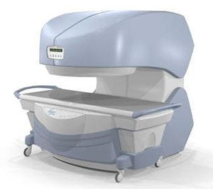
This open C-shaped MRI system eases patient comfort and technologist maneuverability. This low cost scanner is build for a wide range of appli cations. The Virgo™ patient table is detachable and moves on easy rolling castors. Able to accommodate patient weights up to 160 kg, the tabletop has a range of motion of 30 cm in the lateral direction and 90cm in the longitudinal direction. Images generated with this scanner can only be viewed (without data loss) on Millennium's proprietary viewing software.
Device Information and Specifi cation CLINICAL APPLICATION Whole body Head, Body, Neck, Knee, Shoulder,
Spine, Wrist, Breast, Extremity, Lumbar Spine, TMJ
IMAGING MODES Localizer, single slice, multislice, volume, fast, POMP, multi slab, cine, slice and frequency zip, extended dynamic range, tailored RF | | | | | |
| | | | | |
| |
|
ABLAVAR™ (formerly named Vasovist™) is a blood pool agent for magnetic resonance angiography ( MRA), which opens new medical imaging possibilities in the evaluation of aortoiliac occlusive disease (AIOD) in patients with suspected peripheral vascular disease.
ABLAVAR™ binds reversibly to blood albumin, providing imaging with high spatial resolution up to 1 hour after injection, due to its high relaxivity and to the long lasting increased signal intensity of blood.
As with other contrast media: the possibility of serious or life-threatening anaphylactic or anaphylactoid reactions, including cardiovascular, respiratory and/or cutaneous manifestations, should always be considered.
WARNING:
NEPHROGENIC SYSTEMIC FIBROSIS
Gadolinium-based contrast agents increase the risk for nephrogenic systemic fibrosis (NSF) in patients with acute or chronic severe renal insufficiency (glomerular filtration rate less than 30 mL/min/1.73m 2), or acute renal insufficiency of any severity due to the hepato-renal syndrome or in the perioperative liver transplantation period.
See also Cardiovascular Imaging, Adverse Reaction, Molecular Imaging, and MRI Safety.
Drug Information and Specification
NAME OF COMPOUND
Diphenylcyclohexyl phosphodiester-Gd-DTPA, gadofosveset trisodium, MS-325
T1, predominantly positive enhancement
20-45 mmol-1sec-1, Bo=0,47T
PHARMACOKINETIC
Intravascular
CONCENTRATION
244 mg/mL, 0.25mmol/mL
DOSAGE
0.12 mL/kg, 0.03 mmol/kg
DEVELOPMENT STAGE
FDA approved
DO NOT RELY ON THE INFORMATION PROVIDED HERE, THEY ARE
NOT A SUBSTITUTE FOR THE ACCOMPANYING
PACKAGE INSERT!
Distribution Information
TERRITORY
TRADE NAME
DEVELOPMENT
STAGE
DISTRIBUTOR
USA, Canada, Australia
ABLAVAR™
Approved
| | | |
• View the DATABASE results for 'ABLAVAR™' (3).
| | |
• View the NEWS results for 'ABLAVAR™' (1).
| | | | | | Further Reading: | Basics:
|
|
News & More:
| |
| |
| | | Searchterm 'Cation' was also found in the following services: | | | | |
| | |
| |
|
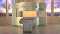
From Hitachi Medical Systems America Inc.;
the AIRIS II, an entry in the diagnostic category of open MR systems, was designed by Hitachi
Medical Systems America Inc. (Twinsburg, OH, USA) and Hitachi Medical Corp. (Tokyo) and is manufactured by the Tokyo branch. A 0.3 T field-strength magnet and phased array coils deliver high image quality without the need for a tunnel-type high-field system, thereby significantly improving patient comfort not only for claustrophobic patients.
Device Information and Specifi cation
CLINICAL APPLICATION
Whole body
QD Head, MA Head and Neck, QD C-Spine, MA or QD Shoulder, MA CTL Spine, QD Knee, Neck, QD TMJ, QD Breast, QD Flex Body (4 sizes), Small and Large Extrem., QD Wrist, MA Foot and Ankle (WIP), PVA (WIP)
SE, GE, GR, IR, FIR, STIR, FSE, ss-FSE, FLAIR, EPI -DWI, SE-EPI, ms - EPI, SSP, MTC, SARGE, RSSG, TRSG, MRCP, Angiography: CE, 2D/3D TOF
IMAGING MODES
Single, multislice, volume study
TR
SE: 30 - 10,000msec GE: 20 - 10,000msec IR: 50 - 16,700msec FSE: 200 - 16,7000msec
TE
SE : 10 - 250msec IR: 10 -250msec GE: 5 - 50 msec FSE: 15 - 2,000
0.05 sec/image (256 x 256)
2D: 2 - 100 mm; 3D: 0.5 - 5 mm
Level Range: -2,000 to +4,000
POWER REQUIREMENTS
208/220/240 V, single phase
COOLING SYSTEM TYPE
Air-cooled
2.0 m lateral, 2.5 m vert./long
| | | |
• View the DATABASE results for 'AIRIS II™' (2).
| | | | |
| | | | | |
| |
|
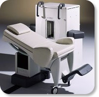
Developed by GE Lunar; the ARTOSCAN™-M is designed specifically for in-office musculoskeletal imaging. ARTOSCAN-M's compact, modular design allows placing within a clinical environment, bringing MRI to the patient. Patients remain outside the magnet at all times during the examinations, enabling constant patient-technologist contact. ARTOSCAN-M requires no special RF room, magnetic shielding, special power supply or air conditioning.
The C-SCAN™ (also known as Artoscan C) is developed from the ARTOSCAN™ - M, with a new computer platform.
Device Information and Specifi cation
CLINICAL APPLICATION
Dedicated extremity
SE, GE, IR, STIR, FSE, 3D CE, GE-STIR, 3D GE, ME, TME, HSE
SLICE THICKNESS
2D: 2 mm - 10 mm;
3D: 0.6 mm - 10 mm
4,096 gray lvls, 256 lvls in 3D
POWER REQUIREMENTS
100/110/200/220/230/240V
| | | |
• View the DATABASE results for 'ARTOSCAN™ - M' (3).
| | | | |
| | | | |
| | | |
|
| |
 | Look
Ups |
| |


